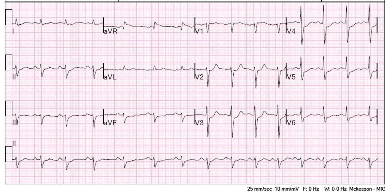
This is a 41 year old male with severe asthma who barely avoided intubation. His troponin I returned at 0.130 ng/mL, and here is his ECG:
 |
||
| There is sinus tach with anterior T-wave inversion of the same morphology as the previous cases of PE which I have posted (see link below). There is no TW inversion in lead III, so this is not pathognomonic. Whether or not there is T-wave inversion, this ECG could be due to PE or any cause of right heart strain. |
Here are some cases of pulmonary embolism.
This is to illustrate that these T-wave inversions are due to acute right heart strain, which is caused by many etiologies other than PE, including but not limited to acute severe asthma and acute pneumonia. Hypoxia causes pulmonary vasoconstriction (pulmonary hypoxic vasoconstriction) which puts strain on the right heart.
Pulmonary embolism was ruled out.