
This 55 year old woman presented with chest pain:
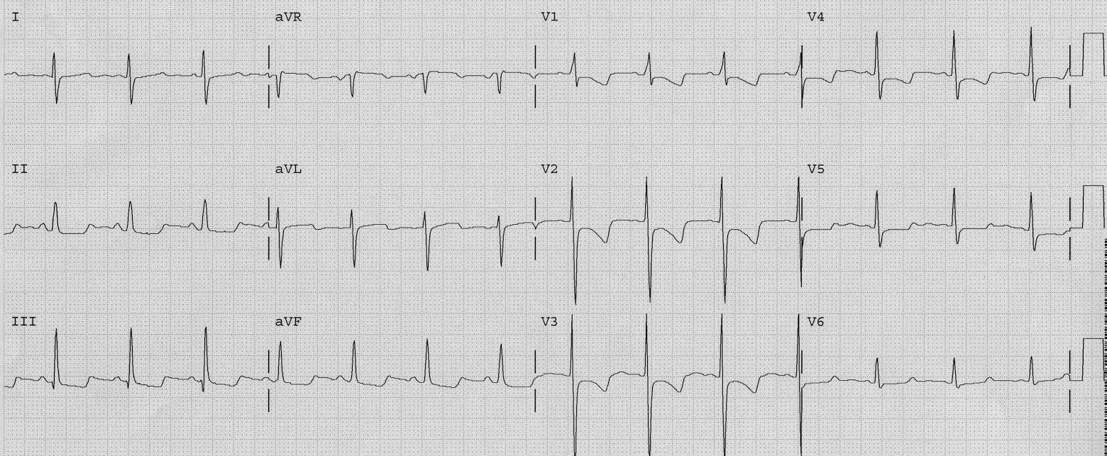 |
| There is sinus rhythm. There is ST depression and there are negative T-waves in V1-V4. There is also STE in aVL, with reciprocal ST depression in II, III, and aVF. Looks like acute posterolateral MI, no? See below. |
No, it is not acute MI. Notice that there is a prominent S-wave in lead I (right axis deviation) and a large R-wave in V1. This is diagnostic of right ventricular hypertrophy, which is an abnormality of depolarization (abnormal QRS) that results in secondary abnormal repolarziation (ST-T wave). These negative T-waves and abnormal ST segments are entirely due to RV hypertrophy.
The physician recognized the abnormal QRS and the appropriate ST-T changes and did not activate the cath lab or initiate anti-ischemic or antiplatelet therapy other than aspirin.
The patient ruled out for MI. Echocardiogram showed RV hypertrophy and pulmonary hyptertension.
EMS 12-lead has a great discussion of pulmonary pressures and ECG patterns, and they show another example of this.