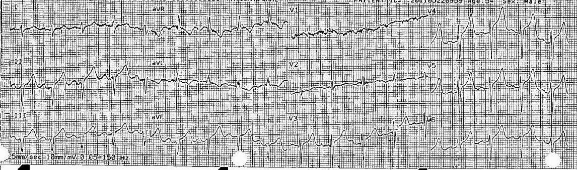
This was sent to me by a reader from India (Thank you, Rama Krishna). The patient presented recently (age unknown). He had a history of DM, HTN, COPD, and previous anterior wall MI.
Here is his presenting ECG:
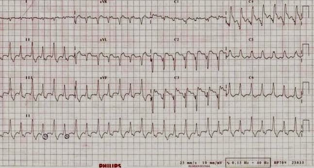 |
The answer is below:
It is ventricular tachycardia with 1:1 retrograde conduction to the atria and also with electrical alternans. The small drawn circles, which appear to be p-waves, are not actually the p-waves. Rather, the small negative deflection following these circles (narrow black arrows along the lead II rhythm strip at the bottom), are retrograde p-waves. They are negative because of the retrograde conduction. The alternate QRS has a negative p-wave that is buried in the negative T-wave (green arrow). These p-waves are upright in aVR (see the blue arrows, and red arrow for the alternate QRS) Also, the retrograde p-wave can be seen in V1 (purple arrow).
As for repolarization, there is ST elevation in V2-V4 (thick black arrow) diagnostic of MI. It looks like it is old MI, not acute (see discussions of LV aneurysm on other posts). Of course, these discussions apply to normal conduction, not normally to ventricular tachycardia. But in this case it is fair to say that the ST segments are a result of MI [almost certainly old MI (which also fits the patient’s history), possibly acute] rather than being due to the abnormal QRS.