Dr. Smith's ECG Blog

Instructive ECGs in Emergency Medicine Clinical Content

Associate Editors:
— Pendell Meyers & Ken Grauer (2018)
— Jesse McLaren & Emre Aslanger (2022)
— Willy Frick (2024)

editors

Dyspnea, Chest pain, Tachypneic, Ill appearing: Bedside Cardiac Echo gives the Diagnosis

A male in his 60s presented complaining of dysnpea and chest pain.  He was tachypneic and ill appearing.

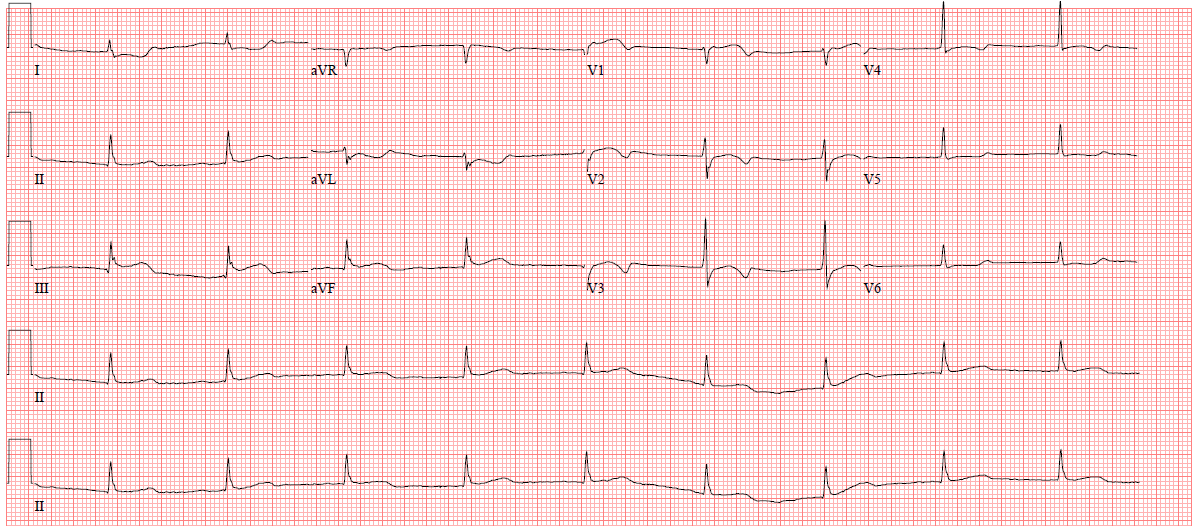
I was texted this ECG.  The physician worried about PE mostly, but also MI:

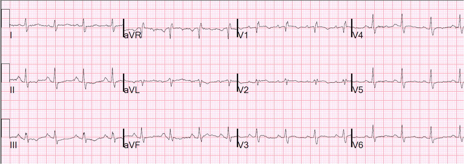
My response was: “incomplete RBBB.  No evidence of acute MI.  Certainly could be PE.  What does the cardiac echo show?”

The bedside echocardiogram showed a serpiginous echogenic structure in the right atrium that was also flowing in and out of the right ventricle.  The RV was enlarged.

I am sorry that I had to take this video down because my colleague wants to submit it to an academic journal.  I have left the still frame in.

Still frame of the video: Blue circle show thrombus snaking throughout right atrium and right ventricle.

The patient was given IV tPA and rapidly improved.  A subsequent ultrasound showed no more thrombus.

Previous Article

COPD exacerbation, what do the ECG and bedside echo show?

Next Article

K. Wang Lecture: the Beautiful World of Electrocardiography. One hour.

View Comments (20)
  1. Hi Dr. Smith,

    Regarding V1, do you interpret a subtle rSR' or an abnormal Q wave?

    -DaveB

  2. There is QR from RBBB with Q-wave, probable old MI, but no acute MI

  3. Interesting case. Silent ECG. Altough this is an awesome type A thrombus this is not the more frequent picture seen (4-8 % PE). Abstracting the thrombus… is still is a PE?…yes, because dilated right chambers, mc connel sign (really evident) and pressure overload findings.
    In other way…has this patient DVT signs and ultrasound confirmation?

  4. Is it possible to have such right atrial thrombus after cardiac arrest.. I remember came across cpr case , we found such echogenic moving mass during pulse check .. could it be due to stasis? After pulse regained, the mass disappeared

  5. Alenia,

    First, I don't see the Q-wave.
    Second, it does not apply in RBBB or incomplete RBBB.
    If it did, 8% of patients with symptoms compatible with PE who also had PE had S1Q3T3, and 2.5% of such patients without PE had it. So there is some utility, but not a lot.
    Steve

  6. ECG is not really silent. It is just not very specific. This was a new incomplete RBBB from 2 weeks prior, which is a typical finding for acute right heart strain. There were clinical signs of DVT but no US confirmation.
    What is McConnel sign?
    Steve

  7. Thrombus due to stasis from cardiac arrest would not be a long, snake-like thrombus (a cast of the lower leg veins), but rather more like the shape of the ventricle.

  8. Laxman Dubey

    q wave in lead V1 only, I don't think it could be due to MI (old). Though Q wave in lead V1-V2 without other abnormalities of MI could be an entirely normal ECG. In one study, QS deflections in both leads V1 and V2 but no other ECG abnormality, only 20% of such patients had clinical CAD or prior MI.

  9. Steve Smith

    Thank you Laxman. Here is Laxman's blog: http://dubeycardiology.blogspot.com/ and here is the paper he is referring to: http://www.ncbi.nlm.nih.gov/pubmed/14731215

    This is also my experience: that many who have no apparent cardiac disease have Q waves in V1 and V2. I am not sure this applies to RBBB also, but it may very well.

    And I see you are from Nepal! A lovely country with lovely people. I have been there 4 times and my wife worked for a year with TB patients near Dankuta.

    Steve Smith

  10. Laxman Dubey

    Dr. Smith thank you for your lovely words. I am a regular follower of your blog, which is very much informative. I would be very glad to invite you again to visit Nepal.

  11. Steve Smith

    Dr. Dubey, thanks so much. I will visit next time I am over there!! Steve

  12. Did you a CT scan for PE?Were you not afraid that after t PA the thrombus will not disappear,but rupture and become a masive saddle pulmonary embolism?Sometimes this can go with cardiac arrest.What about pulmonary thrombectomy?

  13. No CT, just straight to lytics. Your concern is one that is often voice and is real but I think theoretical only. I have never seen this happen nor heard of it happening. Do you know of any papers confirming this?

  14. In the ECG it looks like the P wave in II is large but can't actually count on my comp screen, do you think this represents RA dilatation?

  15. A large P-wave in lead II is due to right atrial hypertrophy and requires chronically elevated RA pressures (pulmonary hypertension). Criteria are 2.5 mm in height. This is less than 2 mm.

Leave a Comment

Your email address will not be published. Required fields are marked *