A 40-something male complained of 3 days of chest pressure. He called 911. He had some pulmonary edema and hypoxia.
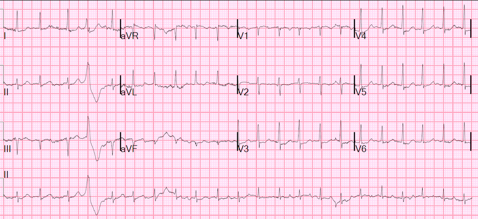
Here is the prehospital ECG:

The computer says ***STEMI***
Based on this and the presence of chest pain, the medics did a prehospital activation of the cath lab.
What do you think?
Interpretation: There is clear atrial flutter. Look at the spikes in V1 at a rate over 300, which are flutter waves. The flutter wave in the inferior leads mimics ST Elevation.
An ED ECG was recorded:

Confirmed Atrial Flutter
Again, notice the flutter waves manifest are sharp spikes in V1
There is 2:1 conduction.
The atrial rate is over 300, with a ventricular response at a bit over 150
The cath lab activation was cancelled and the patient was electrically cardioverted.
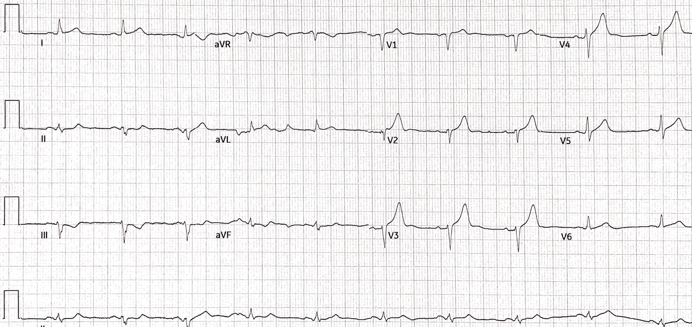
Here is the post cardioversion ECG:

Sinus tachycardia
Look at the P-waves
Notice the very large upright AND inverted portions of the P-wave in lead V1.
Notice also the large P-wave in lead II.
These features indicate right and left atrial enlargement and explain the pronounced size of the atrial flutter spike in V1 during flutter.