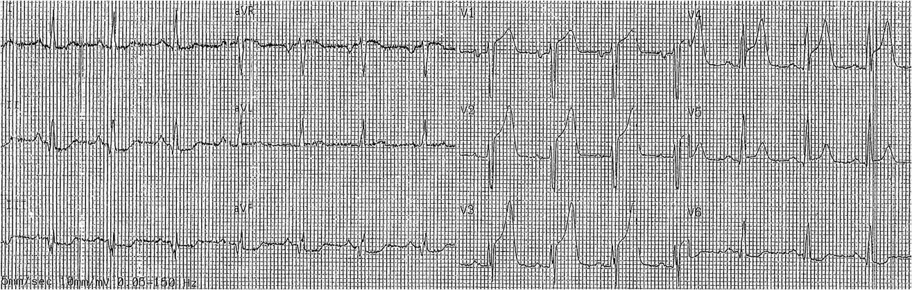
I was shown this ECG and asked if I think it is a STEMI. What do you think?
 |
| Answer Below |
I answered that it does not meet any criteria for STEMI, but that there are hyperacute reciprocal (negative) T-waves in I and aVL and this cannot be anything other than inferior STEMI, especially since the ST depression in V2-V5 suggests concomitant posterior STEMI.
The ECG findings resolved before cath. At cath, there was an 80% hazy thrombotic lesion in the RCA. Thus, this was indeed an acute RCA occlusion. Echo the next day showed an inferior wall motion abnormality.
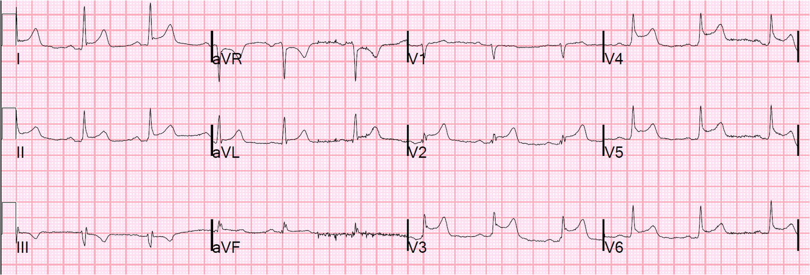
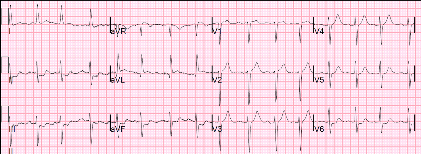
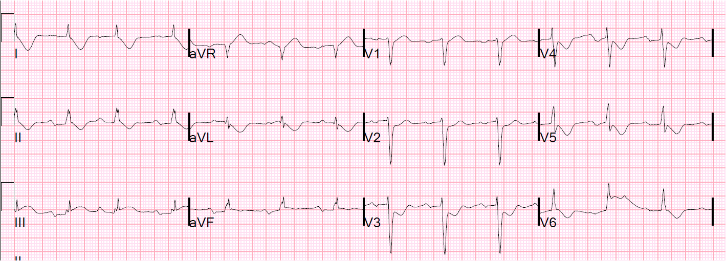
Lesson: Reciprocal ST-T changes are often more pronounced than the ST-T changes overlying the affected myocardium. This is particularly common when there is lateral STEMI with inferior ST depression (see these two cases).
Here is a case in which there are both inferior and reciprocal hyperacute T-waves.