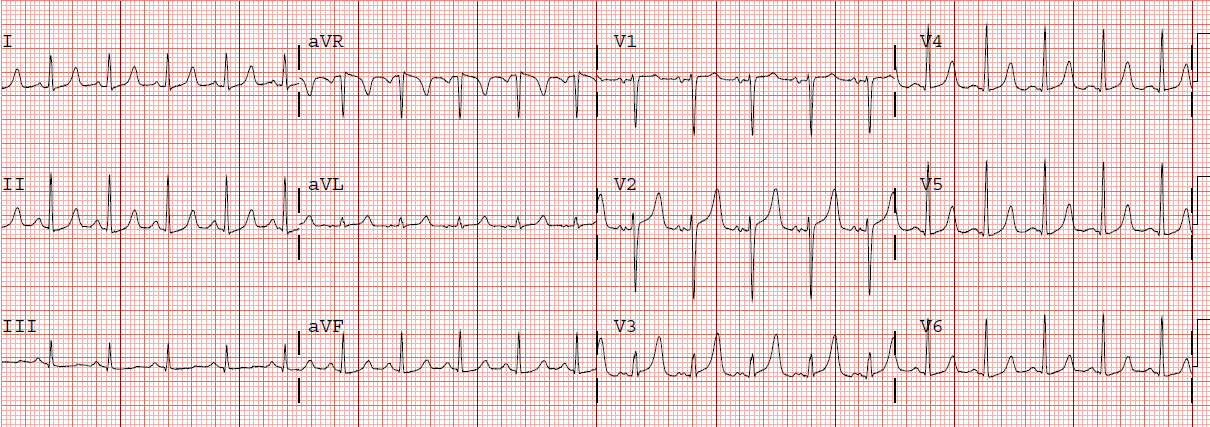
An elderly female with h/o HTN presented with chest pressure. She awoke with bilateral upper sternal chest pressure and tightness, with some SOB, starting at 6 am. Here was her first prehospital ECG at 0623:
 |
| There is sinus rhythm with one PVC. There is subtle ST elevation in aVL and I, with reciprocal ST depression in III and aVF, indicative of a circuflex (or obtuse marginal – OM – branch), or possibly and first diagonal, occlusion. There are also hyperacute T-waves in V4-V6, with some ST elevation, suggesting more widespread STEMI, such as LAD or very large OM or diagonal. The ST elevation is even easier to see in the PVC in lead aVL: it is discordant, as it should be, but out of proportion. |
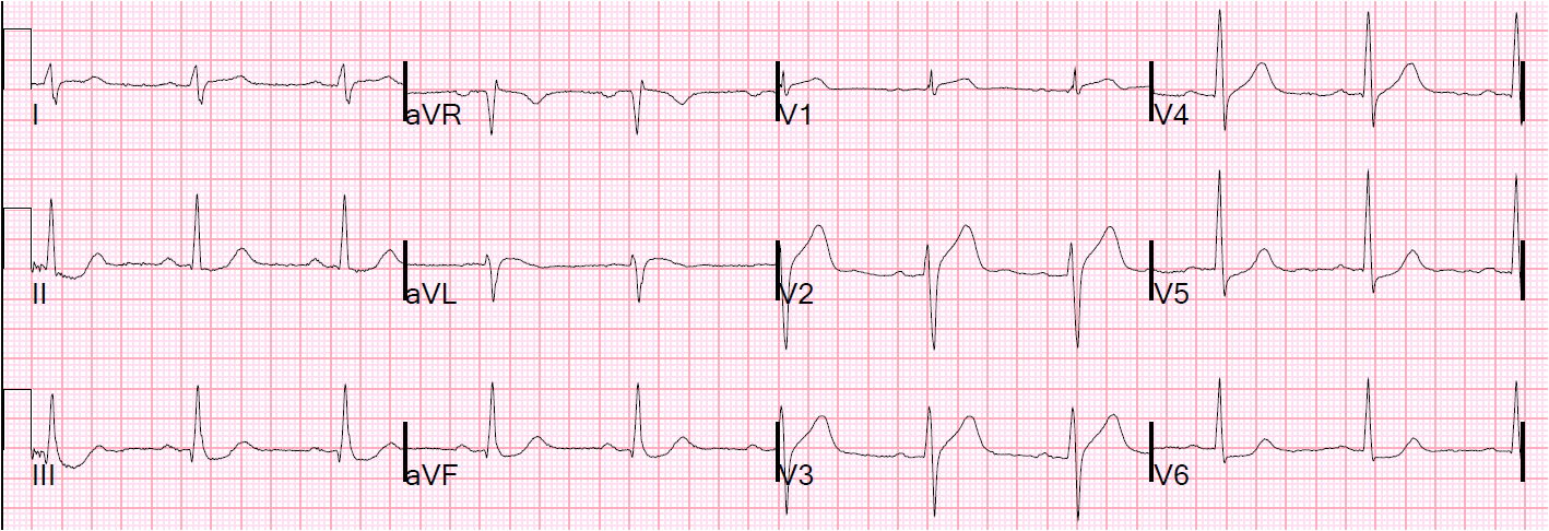
Here was another at 0627:
 |
| This is similar, except that the PVC is now seen in V4-V6, and this time the T-wave appears more hyperacute in the PVC than in the normal beat. |
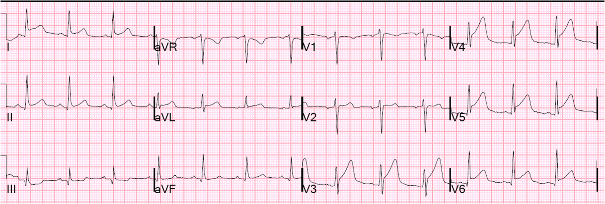
She received nitroglycerin and had near resolution of chest pain. She arrived in the ED at 0639. I looked at the ECG and immediately activated the cath lab. We then recorded another at 0651 while waiting for the cath team:
 |
| There is less high lateral STE (aVL), but still some “inferior” ST depression. Interestingly, there is now new ST Depression in V2 and V3, with a new Q-wave in V2. This is suggestive of LAD disease. The T-waves in V4-V6 are much less hyperacute. |
She went to the cath lab immediately and had co-culprit lesions:
1) 99% thrombotic lesion in the proximal segment of a large obtuse marginal, with TIMI-2 flow
2) 99% mid LAD occlusion with TIMI-2 flow.
Both were stented.