A young man had 10 seconds of syncope without a prodrome. He had had it once before. Exam was normal.
This ECG was recorded:
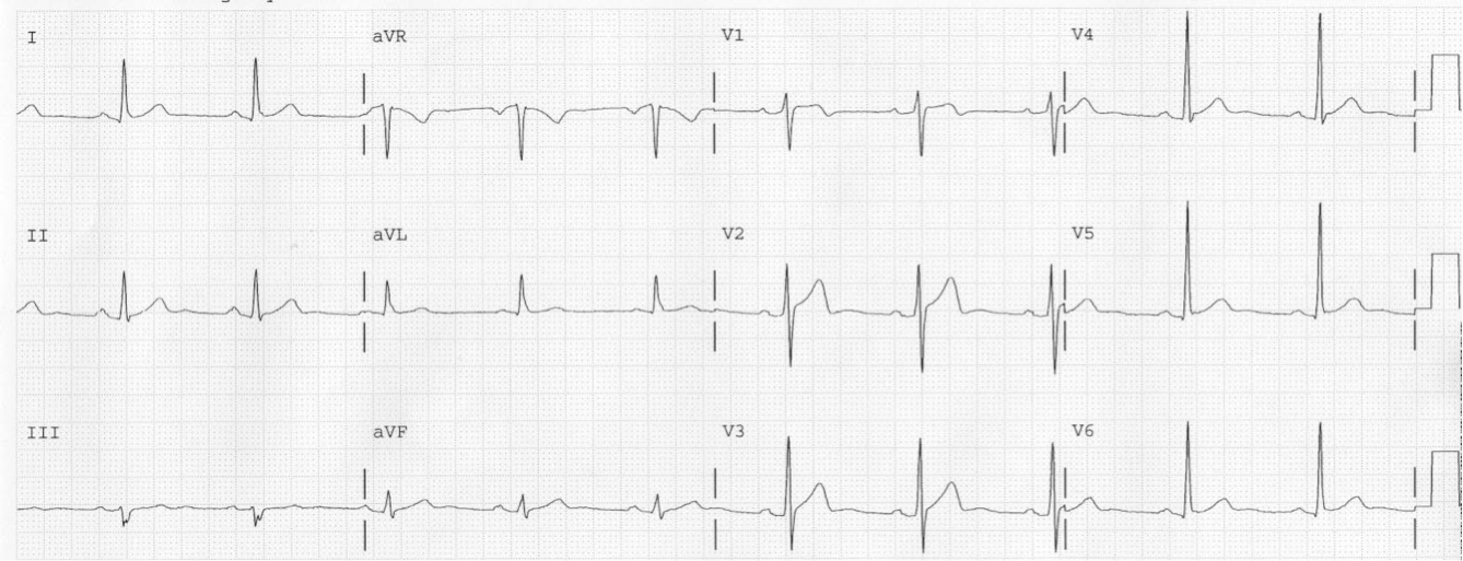
 |
| There is rSr’ in both V1 and V2, with a “saddleback” in lead V2, and the “beta” angle is wide. It meets, or at least nearly meets, criteria for type 2 Brugada. |
Having just written on this topic, I knew that this could be the result of lead placement that is too high.
V1 and V2 should be placed at the 4th intercostal space. Placing them at the 3rd can result in false positives for Brugada.
I asked the tech if she was sure she had placed them correctly.
She returned saying that she had placed them one interspace too high, and handed me this ECG recorded at the right interspace:
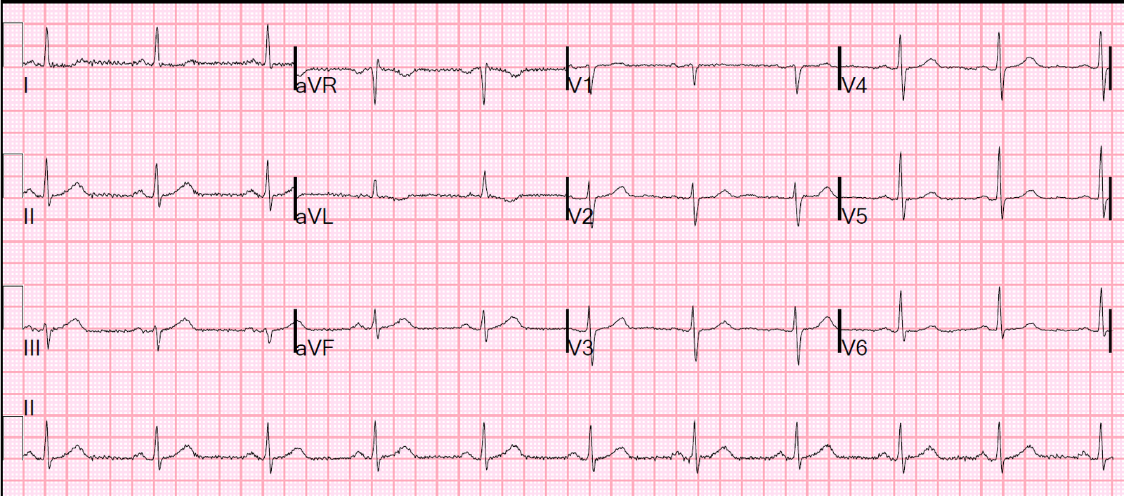
 |
| The rSr’ is gone and the saddle is gone as well. |
Learning point:
Beware lead placement in the diagnosis of right ventricular conduction delay (rSr”) and in the finding of Brugada pattern ECG.