This is a repost, but it was only posted as a video presentation on hqmeded: https://vimeo.com/18680419
A patient presented with chest pain:
 |
| What do you see? (Sorry for the obscured V6. It doesn’t show anything) |
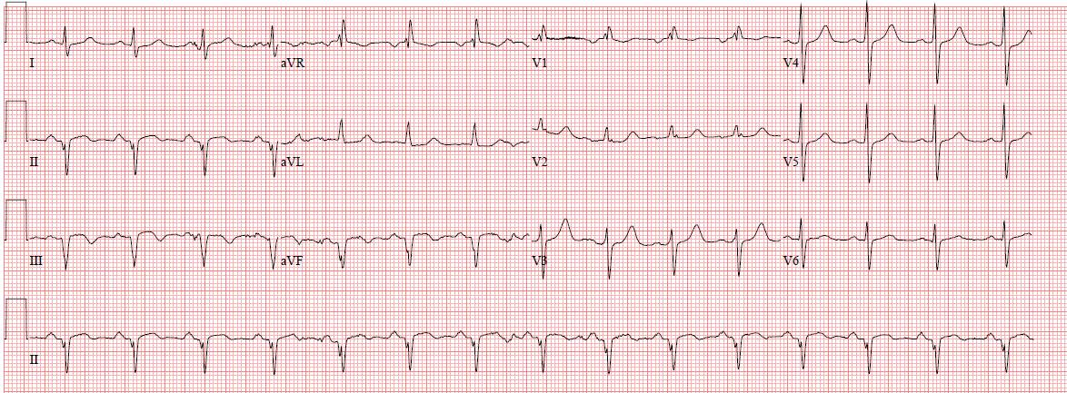
There is an obvious inferior STEMI. The cath lab was activated. I saw the ECG later and called the interventionalist to hear the outcome. He said there was a mid-RCA occlusion. I said, “That is strange, because the ECG has ST elevation in V1 and indicates a right ventricular MI.”
He went back to look at the angiogram. He then saw that the culprit was in the proximal RCA, proximal to the RV marginal branch, and that there had, indeed, been an RV MI. The mid-RCA occlusion was an embolus from the proximal RCA. Fortunately, the patient was still on the table and he could go back and place the stent at the correct lesion in the ulcerated plaque in the proximal RCA.
Learning Points:
1. In inferior STEMI, ST elevation in V1 is a very specific finding for right ventricular MI. We have studied this and are near completion of a manuscript on it.
2. In inferior STEMI, it pays to record a right sided ECG.