Written by Pendell Meyers
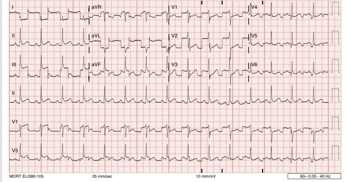
A man in his 40s presented with acute chest pain.
Here is his triage ECG:
What do you think?
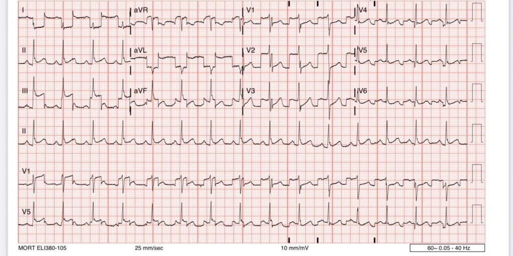
The ECG above shows a dramatic artifact originating from the left arm electrode. Lead II is the only normal/unaffected limb lead present, meaning that there is no significant artifact coming from the electrodes that compose it: RA and LL. All other leads are affected by the artifact which must be coming from the LA electrode.
Why are the precordial leads also affected? I believe this is because Wilson’s central is created from the limb leads and is used as the negative pole of the precordial electrodes to create the precordial leads.
I initially thought the artifact was APTA (arterial pulse tapping artifact), which means that the electrode is being affected by the physical movement associated with being placed near a pulsatile artery (the most classic example is near a patient’s dialysis fistula). In this case, the artifact must be linked 1:1 with the cardiac cycle.
However, Hans Helseth pointed out to me that this is not APTA, because the artifact is not synced to the cardiac cycle. The QRS is seen on different parts of the artifact waveform in multiple leads. So it is not from the cardiac cycle, but rather extrinsic in origin.
the PMCardio Queen of Hearts AI OMI ECG Model was fooled by this one, assessing as “STEMI/STEMI Equivalent”.
It seems that the ECG was not initially understood as artifact. The cath lab was activated.
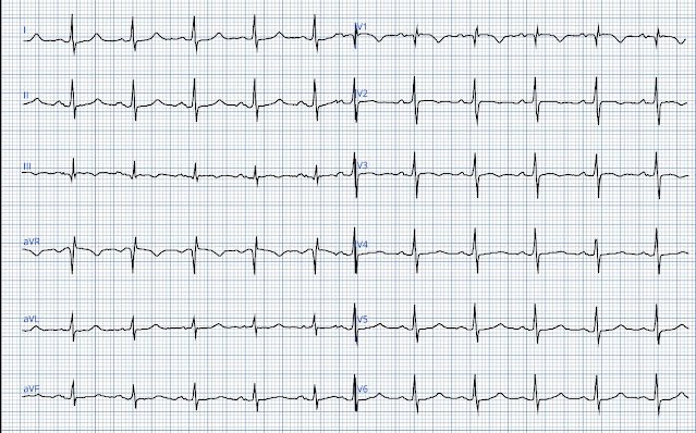
Minutes later, a repeat ECG was performed:
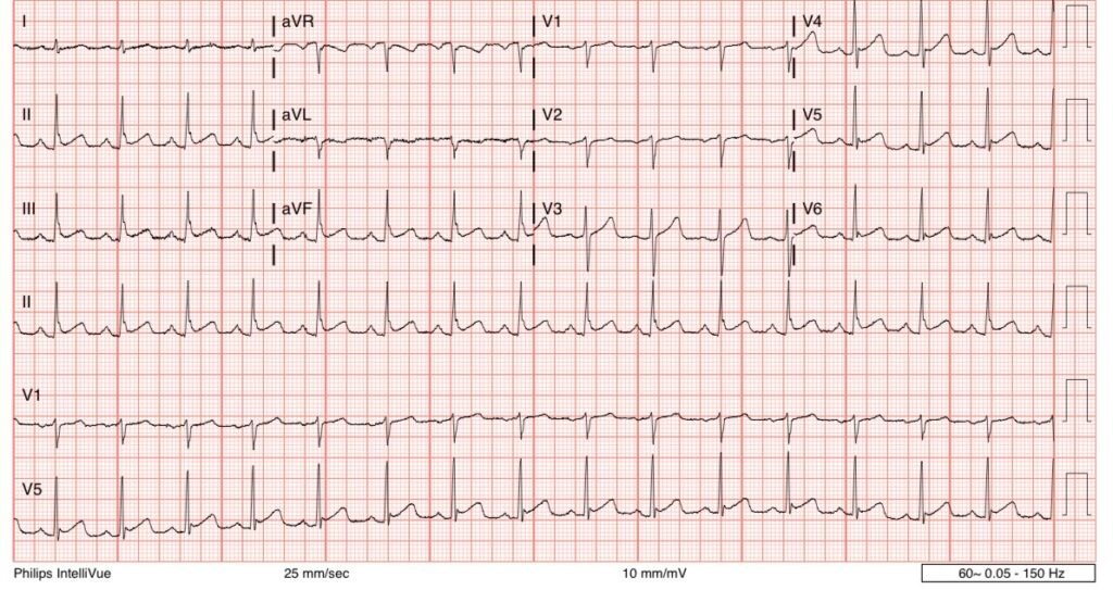
Artifact resolved. Normal sinus rhythm, normal variant STE, no OMI.
The cath lab was deactivated. All troponins were negative. No dangerous cause of chest pain was discovered.
_________________
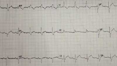
Smith: We have many cases of artifact. Many are Arterial Pulse Tapping Artifact, which often mimics OMI. Here is one.
A 60-year-old diabetic with chest pain, cath lab activated
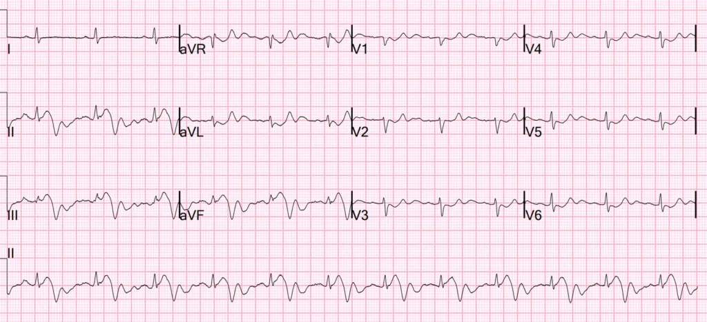
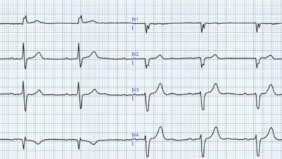
Here is another example of APTA: Are these Hyperacute T-waves?
At this post, there is a good explanation of these artifacts and how to easily recognize them (hint: whenever something looks bizarre, look at leads I, II, and III and see if one of them — only one! — is NOT bizarre)
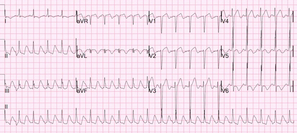
Excellent review of our artifact/APTA related cases by Ken Grauer (See his Comment below):
================================
Links to Examples of ARTIFACT:
Artifact is a reality of clinical practice — so comfort in assessing tracings with artifact is essential. What follows below is my expanding list of technical “misadventures” — most from Dr. Smith’s ECG Blog — some from other sources (Grauer: As I did not previously keep track of these — there are additional examples of artifact sprinkled through Dr. Smith’s ECG Blog that I have not yet included here … — See My Comment below — ).
- The November 8, 2024 post — artifact complicating OMI assessment.
- The June 15, 2024 post ( = Today’s case by Dr. Frick) — re Telemetry artifact that simulates PMVT.
- The May 18, 2024 post — re the effect of baseline artifact.
- The January 15, 2024 post — for an OMI despite lots of artifact!
- The September 15, 2023 post — for PTA (Pulse-Tap Artifact).
- The April 6, 2023 post — excessive baseline artifact misdiagnosed as AFib (instead of sinus rhythm with AV Wenckebach — as in Figure-4 in this post).
- The March 17, 2023 post — for PTA.
- The January 17, 2023 post — for PTA.
- The October 21, 2022 post — for “artifactual VT”.
- The November 10, 2020 post — for PTA.
- The October 17, 2020 post — for a 70-year old woman with “Artifactual VT“.
- The September 27, 2019 post — for the Rowlands & Moore article with the above-noted formulas for recognizing the “culprit” extremity.
- The September 22, 2019 post — intermittent ST-T wave artifact.
- The August 26, 2019 post — baseline artifact.
- The January 30, 2018 post — for PTA.
- Brief review by Tom Bouthillet on some common causes of artifact.
- Additional review of ECG artifacts by Pérez-Riera et al (Ann Noninvasic Electrocardiol 23:e12494, 2018)
- VT Artifact — by Knight et al: NEJM 341:1270-1274, 1999.
- Artifact simulating VFib — CLICK HERE.
- More VT-VFib artifact — CLICK HERE.
- Artifact simulating AFlutter — CLICK HERE.
- Parkinsonian Tremor vs AFlutter — CLICK HERE.
- Left Leg artifact — CLICK HERE.
- Should the cath lab be activated? — CLICK HERE.
- Left Arm artifact — CLICK HERE.
- More PMVT vs Torsades artifact — CLICK HERE.
======================================
MY Comment, by KEN GRAUER, MD (7/23/2025):
NOTE: I have added to Dr. Meyers’ above list of LINKS showing Artifact — as well as to the Techncal Misadventures of Lead Reversals. For easy reference — We have added this as a Tab in the Menu that appears at the top of every page in Dr. Smith’s ECG Blog (As per the Figure below — this Tab is the 4th link under Research & Resources in the Menu at the top of every page).
===================================
— Where to find the “Technical Misadventures LINK in the Top Menu! —
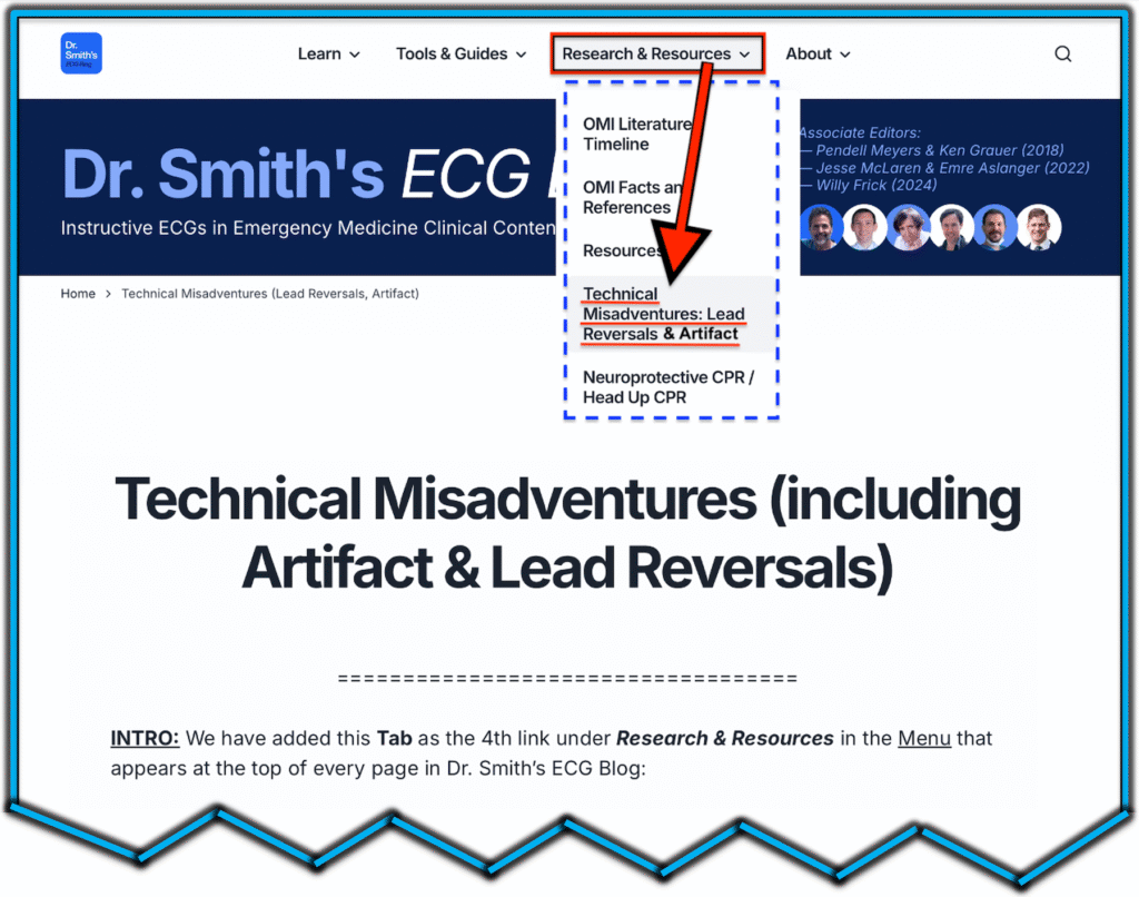
All-too-often lead reversals, unsuspected artifact, and other “technical misadventures” go unrecognized — with resultant erroneous diagnostic and therapeutic implications.
- In the hope of facilitating recognition of these cases — we are developing an ongoing listing on this page with LINKS to examples that we have published in this ECG Blog.
- NOTE: We welcome additional input from our readers! If you have cases to share with us for potential publication — Feel free to email them to me — Ken Grauer, MD (ekgpress@mac.com ).
===================================

