A young man presented after a syncopal episode. He felt fine in the ED. He had no previous cardiac history, but stated that he does have an abnormal baseline ECG.
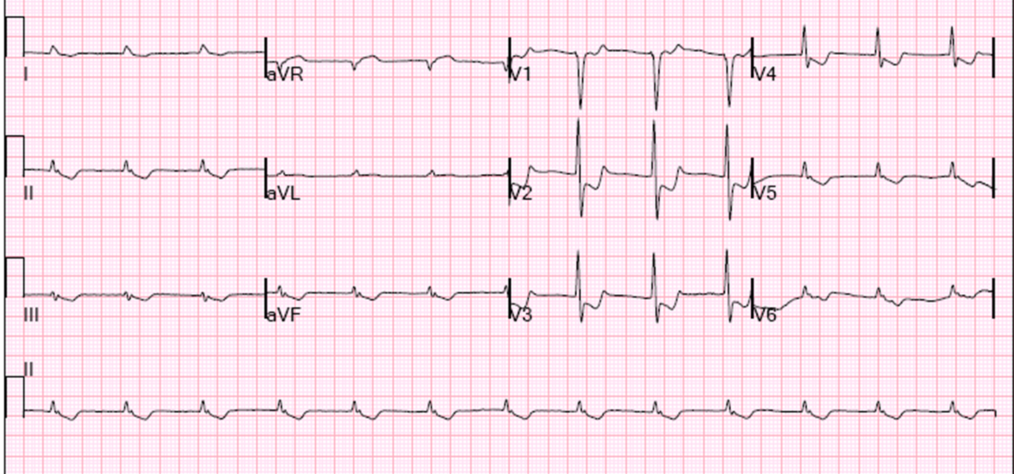
Here is his ED ECG:
 |
| There is a hyperacute T-wave in lead III which is far larger than a small QRS. There is reciprocal ST depression and and inverted reciprocally hyperacute T-wave in lead aVL and I. Precordial leads support the diagnosis of STEMI, showing posterior STEMI. |
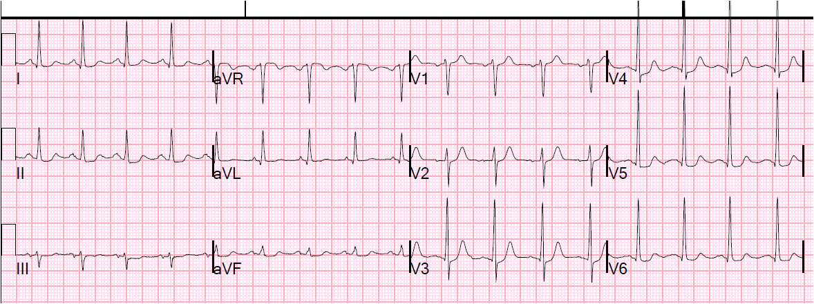
So this young person with syncope and no other symptoms is having a STEMI?
Could it be that this is his abnormal baseline ECG?
NO.
There is no baseline abnormality which looks like this.
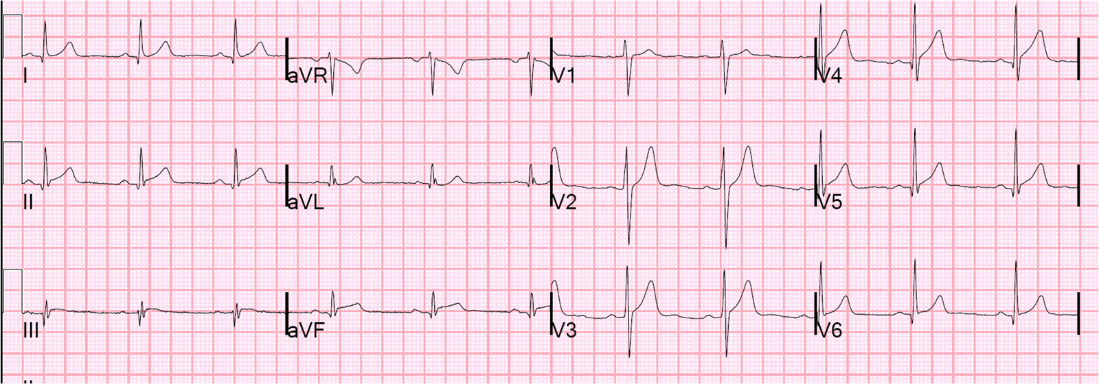
There is a condition of baseline inferior ST elevation (“early repolarization” in the limb leads). We have shown that this never has reciprocal ST depression. Here is the reference:
Bischof J. Thompson RP. Tikkanen J. Porthan K. Huikuri H. Salomaa V. Smith SW. ST-segment depression in lead aVL differentiates benign ST elevation from inferior Acute STEMI. ACEP Research Forum 2012. Annals of Emergency Medicine 60(4 Suppl):S8-S9; October 2012.
The patient was taken for PCI of 100% thrombotically occluded RCA. The etiology of the syncope is uncertain, but probably some dysrhythmia related to the inferior STEMI.
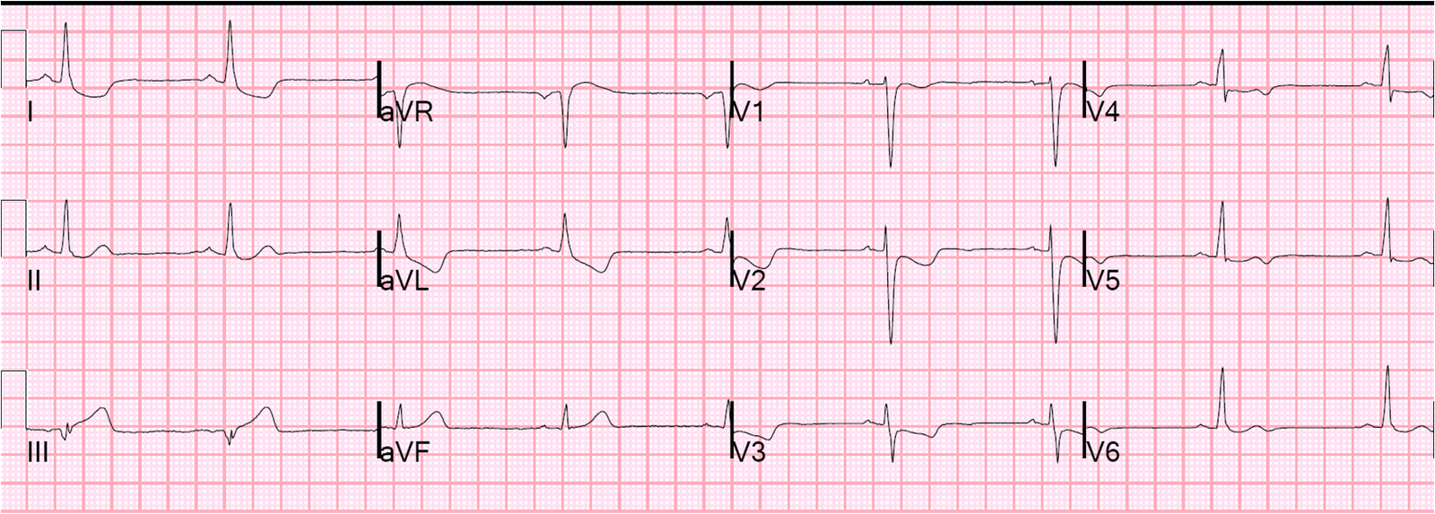
Later, his baseline abnormal ECG was obtained. Here it is.
 |
| This is classic “Benign T-wave Inversion” |
To learn all about Benign T-wave Inversion, read this.