Dr. Smith's ECG Blog

Instructive ECGs in Emergency Medicine Clinical Content

Associate Editors:
— Pendell Meyers & Ken Grauer (2018)
— Jesse McLaren & Emre Aslanger (2022)
— Willy Frick (2024)

editors

A Very Elderly Male with a Fall and no Chest Pain

A very elderly man fell and injured his face.  There is always the possibility of syncope as the etiology of a fall, especially in an elderly person.

I was consulted on his ECG.

He had no chest pain, nor chest pain shortly before presentation.

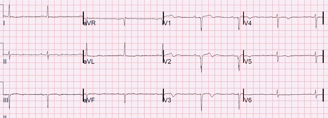
What do you think?
This is what the treating physicians wrote:
“Syncope workup notable shows abnormal EKG with Wellens’ waves in V2 and V3.

My opinion was this:  

“There are no R-waves in V2 and V3, so it is not Wellens’.  Furthermore, it can’t be Wellens’ syndrome even if the ECG is true Wellens’ morphology: Wellens’ syndrome requires that the ECG be recorded after an episode of chest pain that is now resolved.  This patient had no pain at any time.”

I asked:

“Is there a previous ECG for comparison?” (No, there was not)

Continued

“The QS-waves and shallow T-wave inversion are typical of a dense old transmural anterior MI, and may represent an aneurysm of the LV anterior wall.  There could be some acute NonSTEMI superimposed on old MI, but the ECG does not make me worried for LAD acute coronary syndrome and not for acute coronary occlusion.  If these T-waves are due to acute ischemia, then the troponin will be positive.  So just get serial troponins.”

Outcome

The patient ruled out for MI, with all troponins below the level of detection.

A previous echocardiogram was found, which showed

Regional wall motion abnormality-distal septum, anterior and apex, hypokinetic, large.

Previous Article

Bizarre T-wave Inversions in a Patient without Chest Pain

Next Article

Test almost all of your most important ECG rhythm interpretation skills with this case.

View Comments (18)
  1. 1) V1-V3: biphasic T waves with (prominent) U waves ? (Kalium ?)

    2) T-V1 taller than T-V6 (Marriott, 1962)

    3) A bit of QRS-fragmentation in V1-V2 ?

    merci Dr Smith !!

    Al

  2. Thanks for the interesting case (and blog)! Is there a minimum amplitude criteria for the r-wave? From my experience it is common with a small positive deflection as seen in V3 (and V2 to some degree) preceding the QS-complex. This has sometimes
    made me wonder whether there are q-waves or not.

  3. Thanks for an interesting case (and blog)! From my experience it is common with a small positive deflection preceding the Q-wave as is seen in v3 (and v2 to some degree). Sometimes it can be quite big which have made me wonder whether there are q-vawes or not. Do you have any thoughts on how large the deflection would be to still call it a qs-complex?

  4. ECG Interpretation

    GREAT case about a common primary care (as well as emergency care) issue! As per Dr. Smith, this ECG is not acute — but it most definitely suggests prior injury/infarction. In addition to lacking a history of chest pain — this tracing is not “Wellens Syndrome” because the infarction appears to have already taken place, given the QS we see in V1,V2 and no more than very tiny initial r in V3. Moreover, there appears to be initial slurring of the downslope of the S wave in V1,V2 (if not with some notching) — which are features suggesting these complexes do indeed indicate prior scar from previous infarction. What remains unknown from looking at the ECG alone, is WHEN the anterior injury occurred. Potentially it was a couple days earlier (in which case the anterior chest lead ST-T wave changes could represent a “subacute” reperfusion pattern) — or it could have been many days, weeks, months or longer in the past. Here is where History becomes tremendously important! The Framingham studies taught us that as many as 1/3 of all infarcts are “silent” — that is not accompanied by chest pain. Of this number, about half of the 1/3 were totally “silent” (ie, NO symptoms at all) — and the other half had “other” symptoms (ie, not chest pain — but abdominal pain, shortness of breath, general malaise and/or mental status changes). If one carefully asks a mentally competent patient to “Think back” over recent days, weeks, months to anything that might possibly represent an “event” (ie, a couple of days when you just didn’t feel right; perhaps had some chest discomfort or unexplained “flu” symptoms) — you’ll often be rewarded by suggestion in this retrospective history of something that might point to an onset of what in this case was a large anterior infarction that this patient evidently had in the past.

  5. Well, that's possible. Only single fascicle, and even that is very questionable, as there is no q-wave in aVL or r-wave in III.

  6. Concern for hypoK/long QT here? Rate is only about 50 too. Would definitely at least get a K value, if history suspicious for syncope, I'd be worried about TdP.

    Maarten Van Hemelen
    I. M. Resident

    1. Good thought. There are also very large U-waves. And long QT would be of higher risk in the setting of old anterior MI. However, it was almost certainly a mechanical fall. K was normal.

  7. Hi Dr. Smith! Does a patient with Wellen's Syndrome on EKG and a concerning history (recent chest pain assoc with diaphoresis, sob, and near syncopal episode) need emergent cath? Or can they get urgent cath (like the next day)? At my old hospital where I did residency, these people would go straight to cath lab. But at the hospital I'm working at now, they wanted her to go to the floor and have ACS rule out and likely cath in the morning. Thank you for your insight 🙂

Leave a Comment

Your email address will not be published. Required fields are marked *