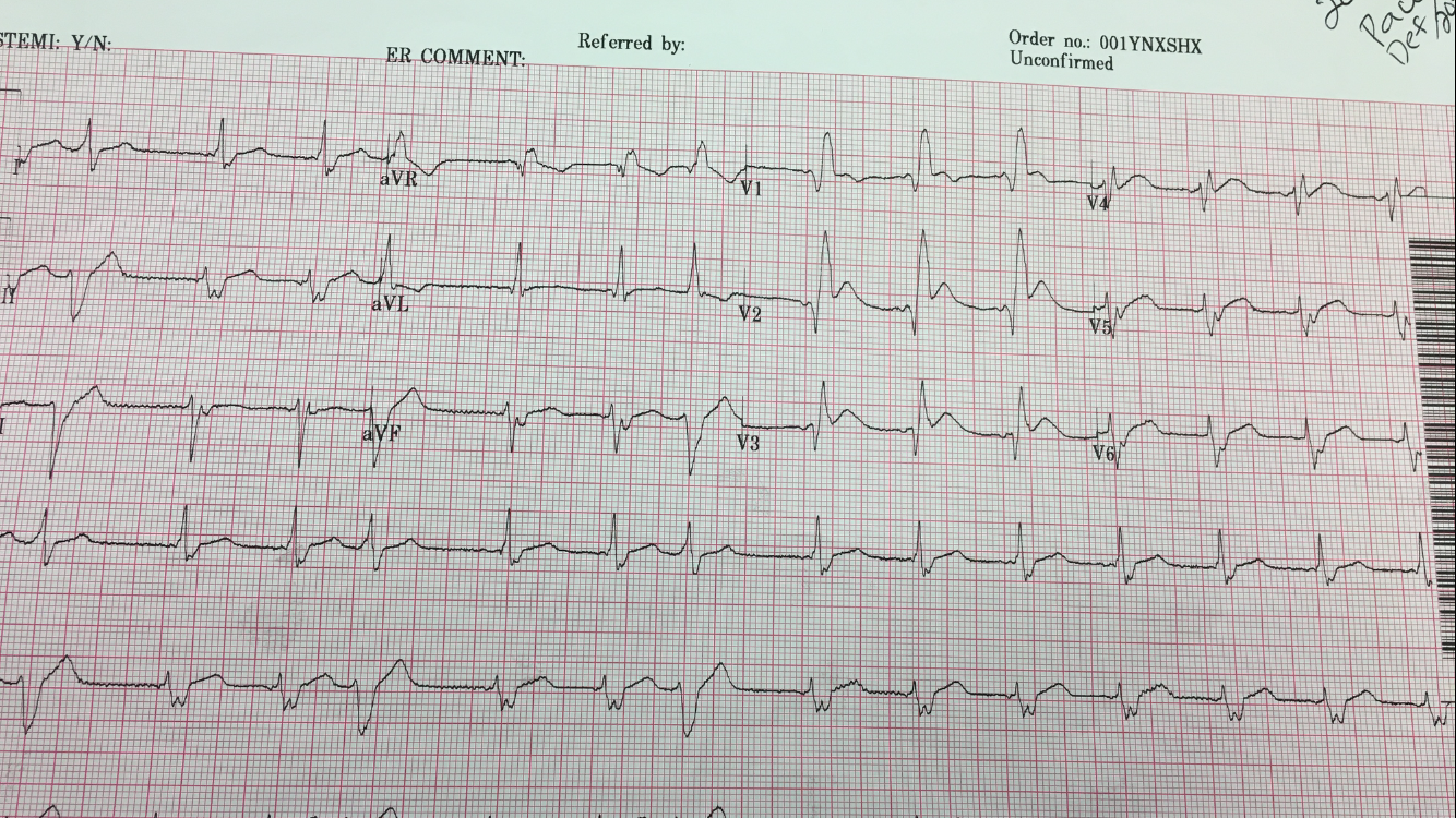
A middle aged woman with few serious medical issues presented with altered mental status, had a generalized tonic clonic seizure, and was found to be hypertensive and tachycardic. An ECG was recorded:
 |
| What is it? The computer measures QRS duration as 126 ms. |
This is a regular, wide complex tachycardia, but not very wide. There are no P-waves. The QRS has 2 alternating morphologies, both of which are right bundle branch block (RBBB) in configuration, but which have different axes. Both complexes have an rSR’ in lead V1 and a wide S-wave in V6.
One might suspect that the higher voltage beats are premature beats, but by my calipers, the intervals are identical. (This is critical to being certain that they are not premature beats.) Similarly, one might suspect every-other-beat pre-excitation, but no delta waves are seen.
See how the S-wave in lead I alternates small, large, small, large… The large S-wave indicates right axis deviation and appears to be due to alternating left posterior fascicular block, in addition to RBBB.
So this is SVT with alternating aberrancy: RBBB, then RBBB + LPFB, then RBBB, then RBBB + LPFB…..
The question of electrical alternans came up. SVT frequently has electrical alternans but, unlike in sinus tach, it is not correlated with effusion or tamponade. In this case, it is not the typical SVT electrical alternans — the alternans is due to alternating conduction.
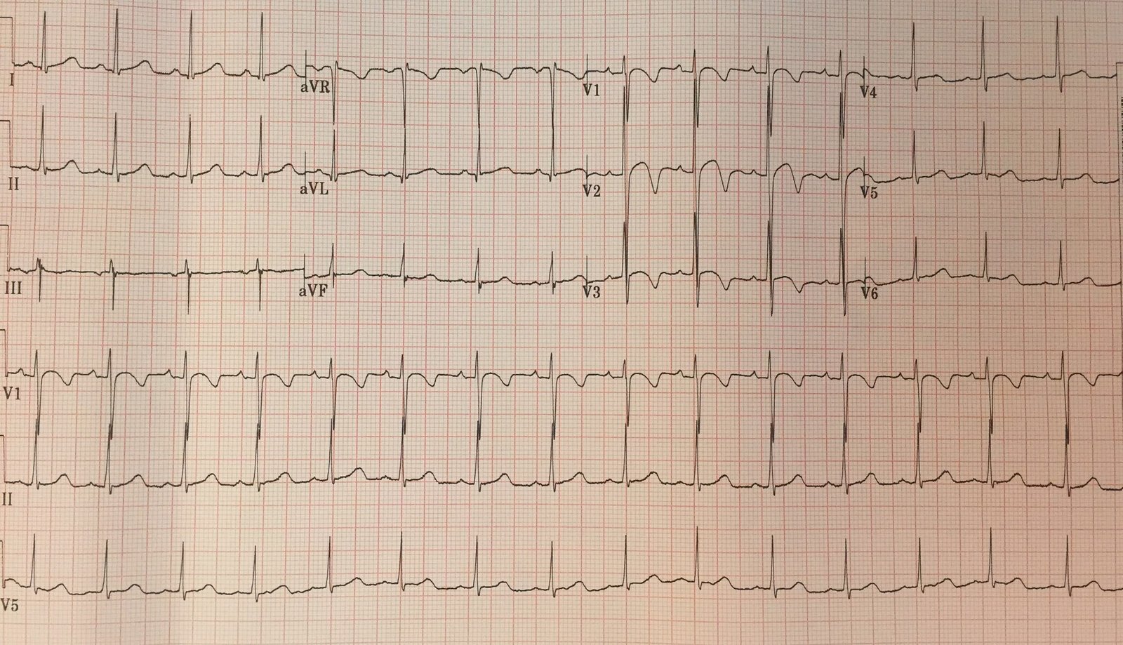
She spontaneously converted:
 |
| Sinus tachycardia with normal conduction.
Normal QRS, narrow, normal axis. This confirms that the SVT caused the aberrancy. ST depression consistent with ischemia. She was found to have a BP of 300/180, and sinus tachycardia at a rate as high as 150. This blood pressure and heart rate were successfully controlled with clevidipine and esmolol. The patient was found on MRI to have “Posterior Reversible Encephalopathy Syndrome” (PRES), which is not always posterior and not always reversible, and which formerly was called “hypertensive encephalopathy.” The extreme hypertension and sinus tachycardia led to suspicion of pheochromocytoma.
An Unusual TachycardiaIs there an alternative explanation? Bidirectional Ventricular Tachycardia may be due to a “ping-pong” effect of alternating VT origin in the left bundle vs. right bundle. It is conceivable that this could be fascicular VT with an alternating origin in the left bundle, then left anterior fascicle, back and forth. Then it would be a bidirectional fascicular VT. I’ve never heard of this before and could not find any literature on it, so it probably doesn’t exist and this hypothesis is pure speculation. See Ken Grauer’s comments on this differential diagnosis, pasted below. As always, it is very incisive!
Ken |