Dr. Smith's ECG Blog

Instructive ECGs in Emergency Medicine Clinical Content

Associate Editors:
— Pendell Meyers & Ken Grauer (2018)
— Jesse McLaren & Emre Aslanger (2022)
— Willy Frick (2024)

editors

An 80-something with weakness, cough, and CP. Should this ECG provoke a Prehospital Cath Lab activation?

An 80-something called 911 for chest pain, generalized weakness, and cough.

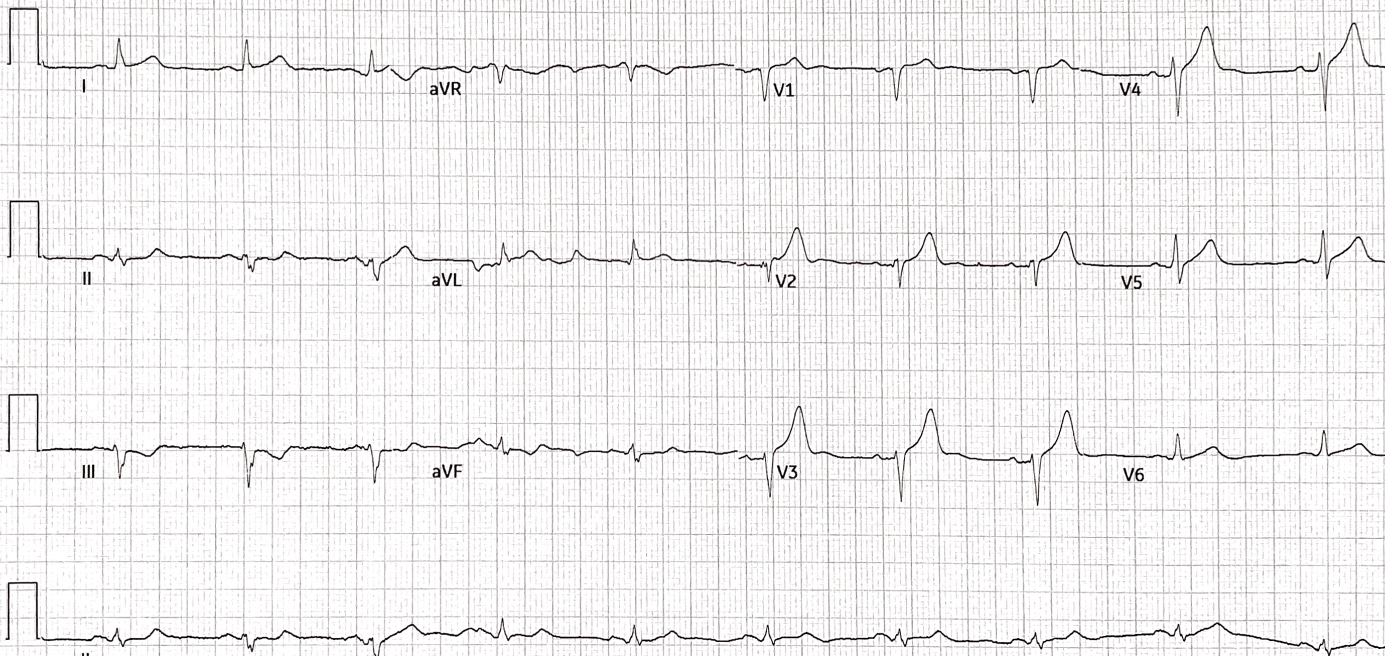
Here is his prehospital ECG:

The medics were worried about this ECG and activated the cath lab

SmithAs in many prehospital ECGs with large voltage, the tracing goes off the image, making assessment of voltage impossible.  Thus, it is impossible to assess the ST Segments and T-waves, which should always be assessed in proportion to the size of the QRS.  I call this “proportionality” and in a non-ischemic ECG, the repolarization should always be proportional to the depolarization.  

Since we can’t see the true size of the QRS, we cannot assess proportionality.

So an ECG like this should not precipitate cath lab activation.

On some machines, one can change the settings of millimeters per millivolt and re-record.  Standard measurements are 0.1 mVolt = 1mm (as in this case), but it can be changed on most ECGs to “half standard” which means that 0.1 mV = 0.5 mm and the QRS complexes then are half sized.

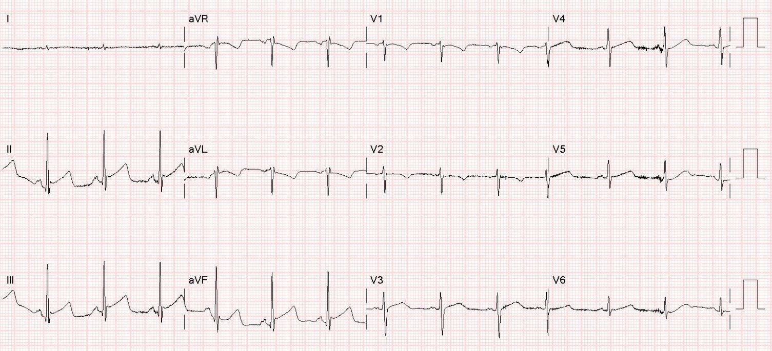
When the patient arrived, an ED ECG was recorded:

Rhythm is uncertain but probably supraventricular at a rate of 100.  

There is RBBB and LAFB and Massive LVH.  

No ST segments are out of proportion to the QRS.

A bedside cardiac POCUS was done, short axis: 

This shows very hypertrophied myocardium

 Long axis

Again, very hypertrophied myocardium

There was no previous ECG on file.

Another ECG was recorded shortly thereafter:

Now P waves are visible in lead V1 

(See biphasic wave approximately 200 ms AFTER the QRS.)  

Thus, there is very prolonged PR interval, at approximately 400 ms.  Then RBBB with LAFB is confirmed.

Thus, this is often called Trifascicular Block.  (Bifascicular block PLUS very prolonged PR interval) and it is associated with progression to complete heart block.  

Read more about “Trifascicular block” here: 

Symptomatic Bradycardia. So-called Trifascicular Block. Occum’s Razor and Hickum’s dictum.

Formal Echo with contrast

–Normal left ventricular systolic function, lower limits of normal; estimated left ventricular ejection fraction is 50%

–Regional wall motion abnormality- mid-inferolateral hypokinesis.

–Marked left ventricular hypertrophy, concentric.

–Normal right ventricular size and function.

The patient turned out to have Covid pneumonia and an elevated hs troponin to 544 ng/L due to type II MI. 

The myocardial diagnosis is hypertrophic cardiomyopathy (HOCM).

Previous Article

Paroxysmal Atrial Fibrillation with RVR, hypotension, volume depletion, good EF, AND pulmonary edema. Strange. Why? What to do?

Next Article

Getting It Right Despite the Wrong Paradigm

View Comments (12)
  1. Thank you for this wonderfull blog.
    One question here.
    Why is this not AIVR on prehospital ecg?
    Thank you very much for your answer!

    1. Good question! Although AIVR is possible, I believe it is actually a supraventricular rhythm (perhaps junctional) with RBBB and LAFB. That is confirmed by the later ECG with P waves AND RBBB/LAFB

  2. Thanks again for a great case, Dr. Smith. I don't tinha I fully understand the concept of disproportional ST segment in the context of RBBB because I see the slight ST segment elevation/depression as disproportional to each preceding QRS. World you mind elaborating on that matter?

    Once again, thank you for the amazing blog, great case and (almost) daily lessons!
    Cheers from Brasil

    1. There is no research on RBBB and LAFB in particular. But when there is IVCD like this, you expect discordant ST Elevation (in the opposite direction from the S-wave). And it should be less than 15%. If over 20-25%, it is definitely ischemic STE.

  3. dr. R.Balaubramanian

    Dr.Smith, Thanks for this great post of excellent educative value(Thursday, April 8th 2021)
    May I indicate 2 pertinent areas for discussion?
    1.Controversies in the diagnostic criteria for diagnosing LVH in the presence of LAFB.
    2.PR interval in the surface ECG – Does it correlate with HV interval (by EPS) linearly ?
    This is an important question since progression to COMPLETE HEART BLOCK would depend on HV
    interval. It is said that PR interval >300 ms is almost always associated with some
    NODAL DELAY as well.
    With regards, Dr.R.Balasubramanian. PONDICHERRY- INDIA

    1. Good point. I am not certain. I do know that the electrophysiologist thought it safe to send the patient home for outpatient workup.

  4. Tall R in aVR, R/S ratio <1 in V6, some evidence of AV dissociation in long Lead II, i thought the rhythm was ventricular origin. Wants to know why it is supraventricular?

    1. VT would have to be much faster. To be ventricular, it would have to be an automatic rhythm because it is so slow. This is possible, but more likely it is supraV with RBBB and LAFB. And the next ECG confirms this.

  5. Your Health and Life Coach

    Thank you dr smith. I am a regular reader of your blog. It’s an interesting case, i thought i saw inferior lateral mi from the first and second ecg. All the best.

  6. Alexey Rukin

    Why are QRS complexes in V5..V6 all NEGATIVE? Shouldn't marked LVH create positive QRS in this direction?

Leave a Comment

Your email address will not be published. Required fields are marked *