Dr. Smith's ECG Blog

Instructive ECGs in Emergency Medicine Clinical Content

Associate Editors:
— Pendell Meyers & Ken Grauer (2018)
— Jesse McLaren & Emre Aslanger (2022)
— Willy Frick (2024)

editors

Atrial Flutter Mimicking ST Depression

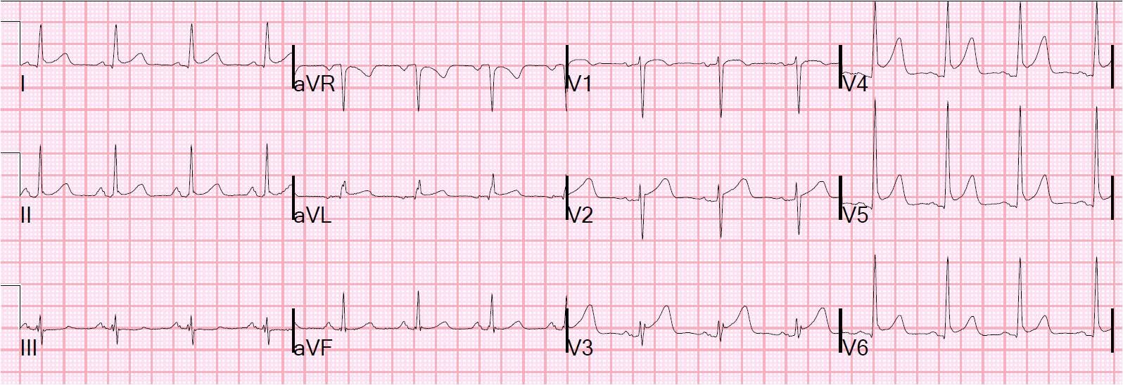
A 65 year old presented with altered mental status and had an intracranial bleed:

One could be fooled into thinking this is sinus tachycardia (with a short PR interval) with diffuse ST depression.  But close inspection reveals flutter waves.  In particular, a totally upright p-wave in V1 is very unusual and should alert you to atrial flutter.  The fluttering baseline accounts for the apparent ST depression, although I cannot rule out some amount of true ischemic ST depression.

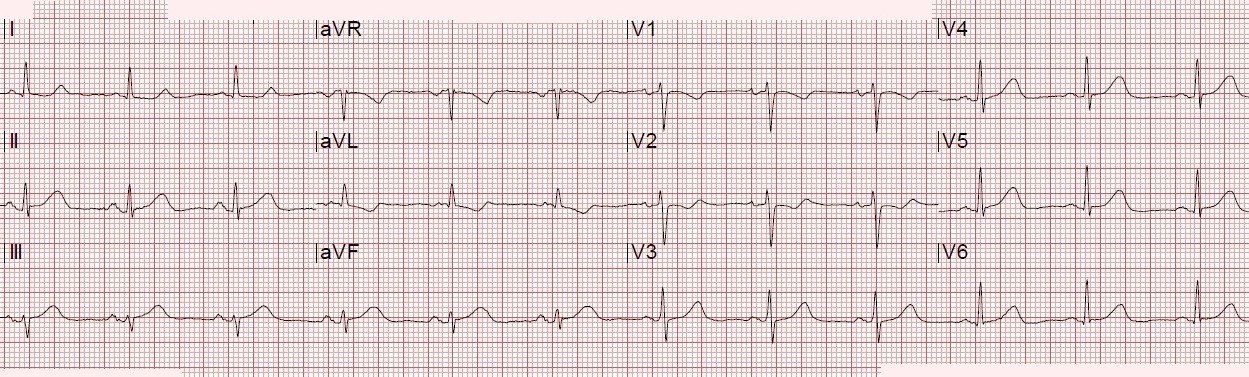
 Here is the ECG after cardioversion:

Now there is sinus.  Interestingly, this one also has an upright p-wave in V1 – so the rule is not universal!
Previous Article

Osborn Waves and Hypothermia

Next Article

Several Cases of ST Elevation from Early Repolarization

View Comments (3)
  1. Dr. Smith,
    the P Wave in Sinus is really upright, but also in II, III there its somewhat biphasic and in aVR slight positiv, isnt it somewhat atypical for Sinus??

  2. O Poder da Eletrocardiografia

    Great Post! The Systematic Approach "Watch your Ps, Qs and the 3 Rs" is useful. The rhythm in first ECG is a regular SVT (Narrow & Regular/SupraVentricular Tachycardia) at ~130/minute without clear sign of sinus P waves. The pattern of ST segment deviation that we see here (ie, ST depression in no less than 7 leads — with ST elevation in aVR & V1-3) is suggestive of subendocardial ischemia. There is LVH(deep S wave in V1 ~28 mm and 30 mm in V2). As per Ken KEN GRAUER the principal differential diagnosis of a regular SVT without clear sign of sinus P waves includes: i) Sinus Tachycardia; ii) Reentry SVT (either AVNRT if the reentry circuit is contained within the normal AV nodal pathway — or AVRT if an accessory pathway is involved); iii) Atrial Tachycardia; or iv) Atrial Flutter. Regularly occurring peak to peak or valley to valley of the flutter waves would be easily appreciated in lead V1 and lead II respectively in Aflutter, but I can't see this in first ECG.I want to know why isn't the other(such as AVNRT or AVRT; Atrial Tachycardia)?. Why AFlutter? Thanks A LOT.

Leave a Comment

Your email address will not be published. Required fields are marked *