See this ECG:
 |
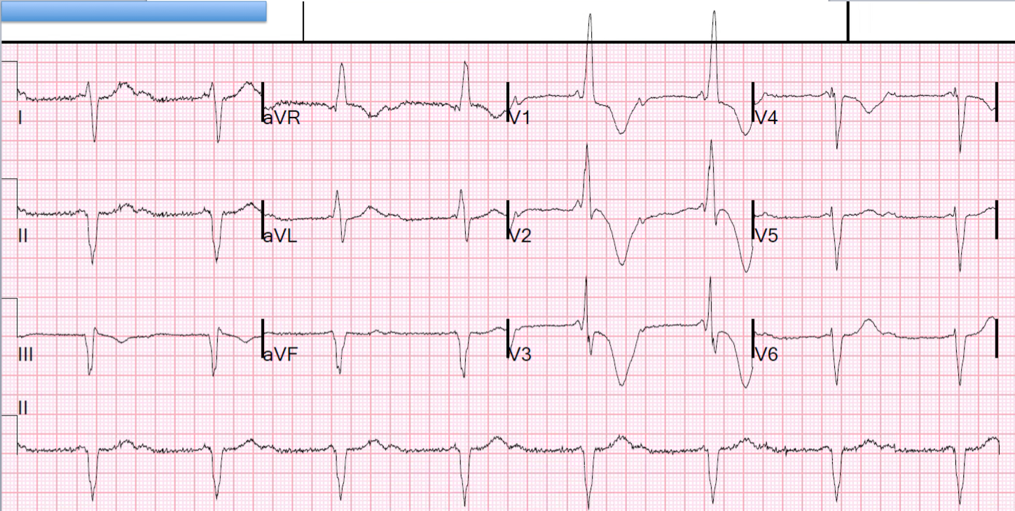
| There is an irregularly irregular rhythm. The Automated interpretation was “Atrial Fibrillation.” What is it? |
Look at the lead II rhythm strip across the bottom. There are clearly sinus P-waves for the first 6 beats, although they speed up.
This change of rate of the sinus node is called “sinus arrhythmia” and is related to vagal tone from inspiration (which increases vagal tone and slows down the rate, but this takes several seconds and this gets out of phase, which means that by the time it is slowing down, the patient is actually expiring).
Then beats 7 and 8 appear and do not show P-waves in lead II. Are they junctional? No! Look above in V1-V3, and you clearly see an atrial beat but of a different morphology (coming from another focus in the atrium, and thus not a sinus beat). This is occurring because the vagal tone is slowing the sinus node so much that a different part of the atrium “escapes,” taking over the pacemaker function.
Beats 9 and 10 also appear to be preceded by subtle atrial activity, but of yet another morphology and thus from yet another focus in the atrium.
So there appear to be at least 3 atrial pacemakers here (3 foci).
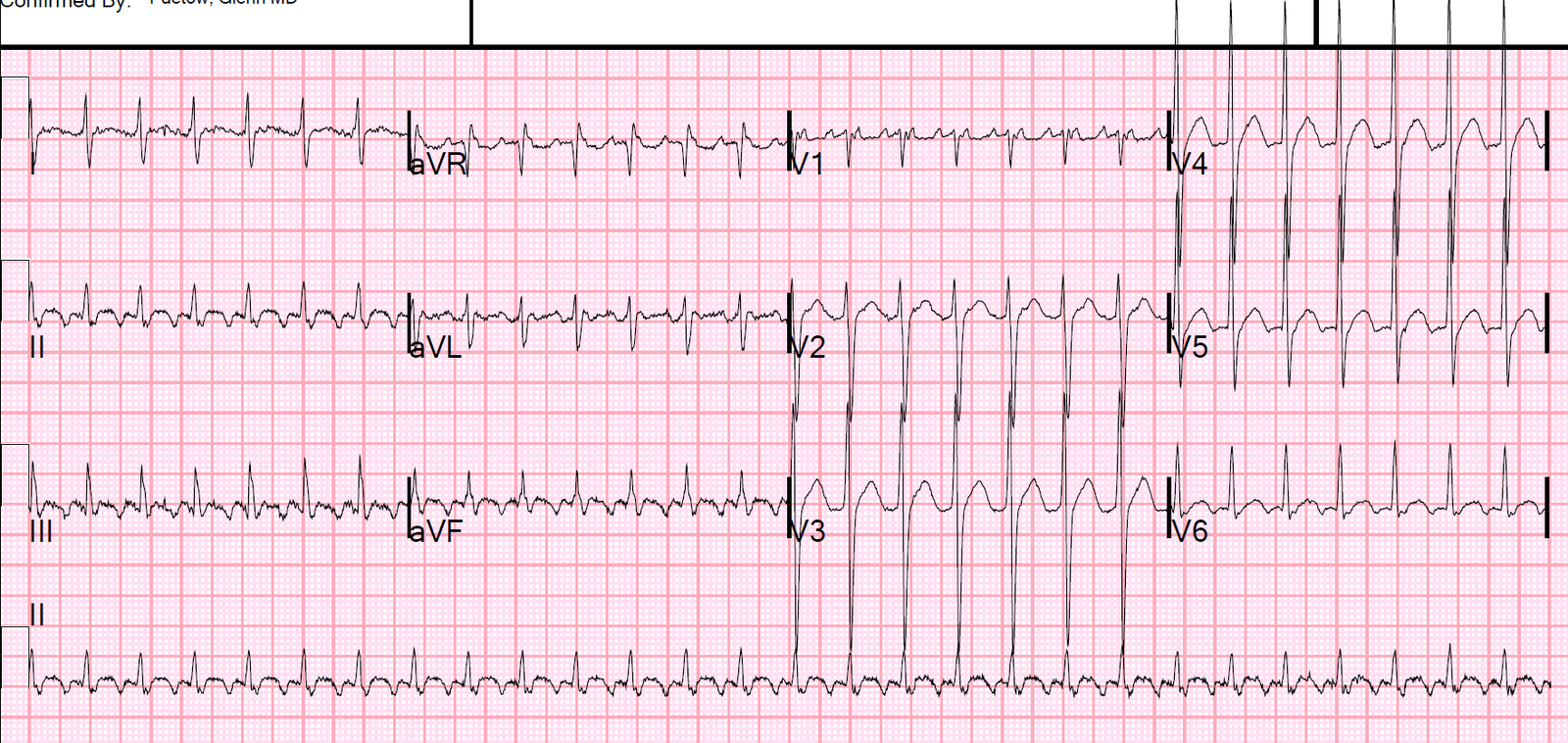
When the rate is tachycardic (greater than 100) and there are at least 3 foci, then it is called multifocal atrial tachycardia (MAT), which is usually associated with COPD. For more on MAT, see this lecture on Narrow Complex Tachycardias from minutes:seconds 23:44 to 26:50.
Since the rate is normal, this is called a Wandering Atrial Pacemaker. It is benign.
2 reasons for an irregularly irregular rhythm in a narrow complex*
1. Multifocal atrial tachycardia
2. Atrial fibrillation
* Sinus arrhythmia appears to be irregularly irregular during the 10 seconds of a 12-lead ECG, but it has a regular pattern to it over more time (speeding up, slowing down, speeding up, slowing down).
Automated interpretations in atrial fibrillation
We compared the Veritas automated interpretation [a widely used algorithm on Mortara machines which is a conventional (if, then; instructional) algorithm] and a new deep neural network algorithm (Cardiologs). We used an expert reference standard, and found that the Veritas had a very large number of false positive reads, more than Cardiologs.(1)