Dr. Smith's ECG Blog

Instructive ECGs in Emergency Medicine Clinical Content

Associate Editors:
— Pendell Meyers & Ken Grauer (2018)
— Jesse McLaren & Emre Aslanger (2022)
— Willy Frick (2024)

editors

Head On Motor Vehicle Collision. ST depression. Myocardial Contusion?

A woman in her 40’s was involved in a head-on motor vehicle collision.  EMS noted a facial droop. On arrival she was hypertensive but other vitals were normal.  She did complain of back and neck pain, chest pain, and SOB.  She also had a facial droop as noted by medics.  This droop was apparently new.

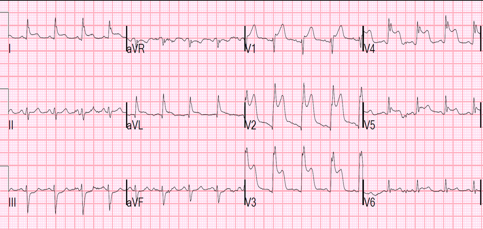
An ECG was recorded:

This shows diffuse ST depression (diffuse subendocardial ischemia) in leads I, II, aVF an V3 to V6, with reciprocal ST elevation in aVR and V1.  This is diagnostic of subendocardial ischemia or injury.  

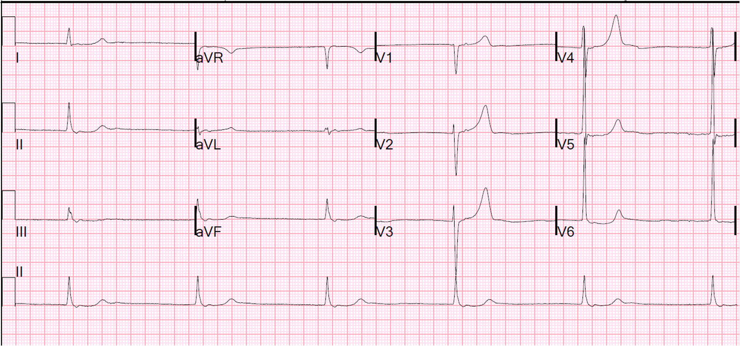
The ECG was repeated 36 minutes later:

The findings are improving

Is this:

1. Myocardial contusion?

2. Stress Cardiomyopathy due to stroke?

3. ACS?

4. Type II (demand ischemia) from stable coronary disease with stress?

5. Aortic
dissection affecting both carotid artery and aortic coronary cusps?

Previous Article

History of Hypertrophic Cardiomyopathy (HOCM), with Tachycardia and High Lactate

Next Article

Is the LAD really completely occluded when there are de Winter's waves?

View Comments (15)
    1. Steve Smith

      when stroke causes these findings, it is generally a very large stroke. This was a very tiny one only causing some facial assymetry.

  1. Dr Magnus Byröd

    Very interesting case. As i have understood it from dr Mattus blog, ST-elevation in aVR would suggest occlusion:
    http://ekgumem.tumblr.com/post/17948970835/lead-avr-deserves-your-respect-episode

    There is also quite a few referenses in the shownotes like Rokos supporting this:
    http://www.ahjonline.com/article/S0002-8703%2810%2900758-1/fulltext

    Am i missunderstanding this, or is it more a case of different opinions? Would be really interesting to hear your thoughts on this!

    1. Steve Smith

      This is a widespread misunderstanding throughout the literature. I am trying to dispel this persistent myth, that in fact is both wrong and makes no sense electrocardiographically.

      Read the last section of my article here. It is on ST elevation in aVR, in both STEMI and NonSTEMI:
      http://link.springer.com/article/10.1007/s40138-012-0003-1#page-2

      Let me know if the link does not work. It should.

      Steve Smith

    2. Dr Magnus Byröd

      Thanks for the link, works fine!

      I guess the most important thing is what you do about these patients. Dr Mattu suggests urgent cath and that you stay of clopidogrel since there is a high chance of CABG, which seems reasonable. Would you send a patient straight to cath lab or try medical treatment first if they presented to the ED with this ECG and complaining of chest pain(without car accident)?

    3. Steve Smith

      I think immediate cath is optimal, but if there are resource limitations, it is acceptable to try medical therapy first: aspirin, heparin, Nitro, GP IIb-IIIa. If ischemia persists, then cath. If chest pain and ST depression resolve, then put on continuous 12-lead ST segment monitoring and cath next day.

    1. Steve Smith

      Not in this case. You should suspect it if there is inferior MI, in which case STE in V1 is due to subepicardial ischemia beneath the V1 lead (of the RV). In this case, there is diffuse subendocardial ischemia, giving an ST depression vector away from lead V1, which causes reciprocal ST elevation in V1 !! (as well as aVR). Confusing, I know. I hope you understand my explanation.

  2. Want to ask.. got 1 patient got intracranial bleeding then when for operation. Come back from operation.. patient's ecg got very wide qrs complexes and tall n wide t wave. The t wave getting taller and taller and more wider compare to the previous ecg. On often got a lot pvc and non sustain vt.. why the t wave behave like that.. and the patient heart is normal.. echo ef 60 %.

  3. I can't understand… STE in AVR and V1, and STD in almost all the others… My first DD was left main occlusion… At least, i think is a powerfull diagnosis to think about…

    1. that is nonspecific and also dependent on lead placement, but could indeed represent old septal MI

Leave a Comment

Your email address will not be published. Required fields are marked *