Dr. Smith's ECG Blog

Instructive ECGs in Emergency Medicine Clinical Content

Associate Editors:
— Pendell Meyers & Ken Grauer (2018)
— Jesse McLaren & Emre Aslanger (2022)
— Willy Frick (2024)

editors

How are these cases related?

I saw these two cases on the same day.

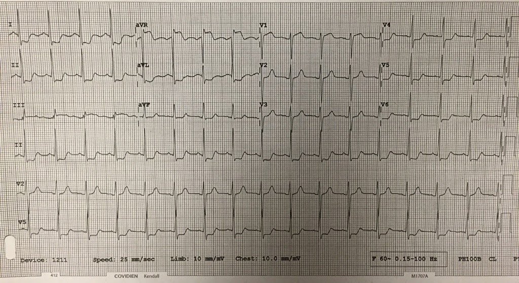
This patient had a GI bleed and a massive transfusion:

What is it?

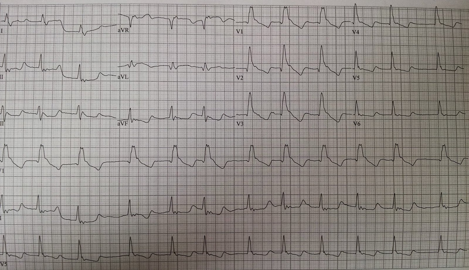
This patient had a history of “frozen shoulders,” and had been treated for this elsewhere for quite a while.  He had been seen in the ED 6 days prior for increased shoulder pain, and was referred back to his orthopedic clinic.  He had this ECG recorded because shoulder pain can be a symptom of ACS:

What do you notice?

The first case has a very long ST segment and thus long QT.  This is classic for hypocalcemia; the ionized calcium was 3.0 mEq/L.  This is a common complication of massive transfusion.  One must be vigilant for hypocalcemia.

The second case shows a very short QT with short ST segment.  The computer measured it at 354 ms.   This was a tipoff to hypercalcemia and so we suspected that this patient had cancer as the etiology of his pain.  A chest x-ray (which we were going to get anyway) confirmed a chest mass.  A chest CT confirmed this and also showed otherwise occult spread to the shoulders.  The ionized calcium was 7.32 mg/dL and the total calcium was 15 mg/dL.

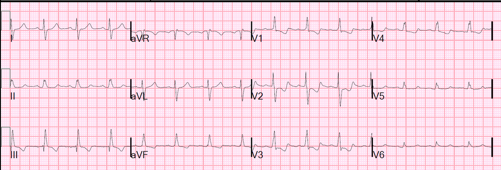
Here was the ECG after normalization of Ca in the second (hypercalcemia) case:

The QTc is now 384 ms
Previous Article

Wide Complex Tachycardia in a 20 something.

Next Article

Interesting Left Main Occlusion....

View Comments (8)
  1. ECG Interpretation

    Hypocalcemia typically produces the "tent sign" at the end of the desert (ie, relatively normal but prolonged ST segment followed by an otherwise unaffected T wave) – which is precisely what the TOP ECG shows. Hypocalcemia often accompanies hyperkalemia – so that T wave at the end of the long ST may be peaked in renal failure patients with both abnormalities.

    ECG-2 is a beautiful example of that short-Q-to-peak-of-T interval seen with hypercalcemia. Having looked for examples of Hypercalcemia on ECG over a period of ~ 3 decades while I was attending (and religiously checking serum Ca++ levels whenever I suspected this finding) – it is not common to see (and be able to recognize) hypercalcemia on ECG in my experience. In those cases in which I could – serum Ca++ levels were almost always quite elevated (ie, ≥13 mg/dL range) – as they were for the above example.

    NICE case!

    1. Olivier,
      My inclination is to attribute it to artifact, of which there is quite a bit on this ECG.
      Steve

  2. Okay, I am stumped but very curious on how these two cases are related. Can you give any tips, Dr. Smith?

  3. Thanks for this, very interesting. I got the long QT but not the short.

    At what point would you say QTc is pathologically short and check the calcium?

    I also thought there are some additional p waves in top tracing, possibly 2:1, but as you say difficult to see with the artifact and no rhythm strip.

Leave a Comment

Your email address will not be published. Required fields are marked *