This was sent by Brandon Stein.
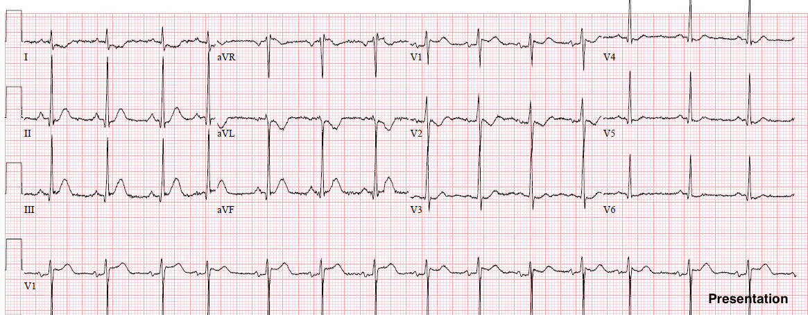
This patient presented with chest pain:
 |
| What do you think? |
There is “inferior” ST depression. When you see this, especially with down-up T-waves, as in III and aVF, it is reciprocal to barely visible ST elevation in aVL. Then look at V2 and V3. Both have hyperacute T-waves, which suggests the LAD. The combination of STE in aVL and V2 is typical of a first diagonal occlusion (the first lateral branch off the LAD).
It was described in the below article. They called it a “mid-anterior” or “mid-anterolateral” infarction without involvement of the septum or apex.
Brandon was very astute and recognized the acute coronary occlusion. The cath lab was activated and there was a first diagonal 100% occlusion.
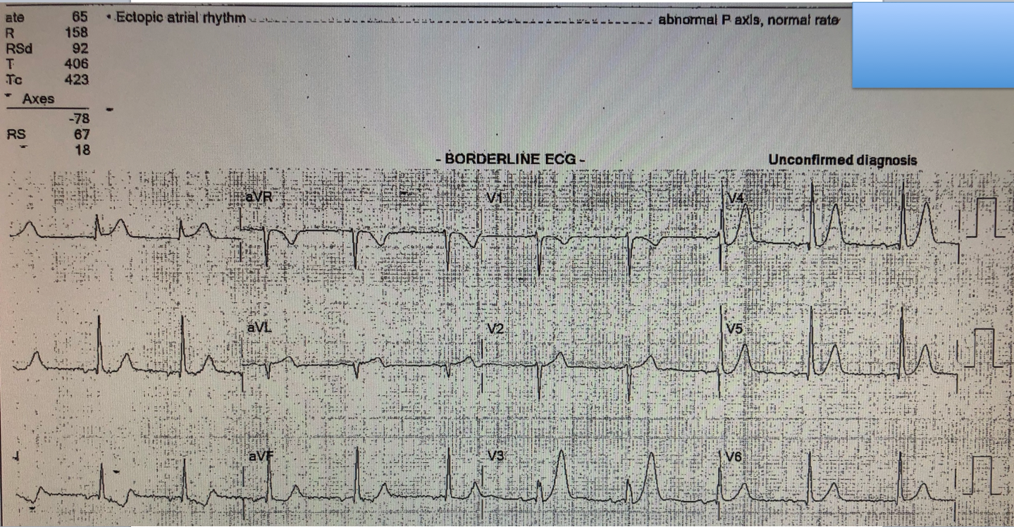
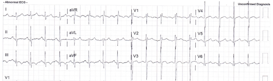
Here is the post cath ECG:

Compare to the presentation ECG:
 |
| The post cath ECG has significantly small T-waves in V2-V5 All of the STE in I and aVL is resolved. All of the inferior ST-T ischemia is resolved |
Peak troponin I was only 1.36 ng/mL, thanks to immediate recognition and rapid reperfusion!!
Learning Points:
1. Coronary occlusion need not have “diagnostic” levels of ST elevation.
2. NonSTEMI frequently needs the cath lab NOW.
3. First diagonal occlusions have their own footprint that is recognizable. Its sensitivity and specificity are really unknown, however. But every time I see it, I recognize it and have not been wrong yet.
Reference