Dr. Smith's ECG Blog

Instructive ECGs in Emergency Medicine Clinical Content

Associate Editors:
— Pendell Meyers & Ken Grauer (2018)
— Jesse McLaren & Emre Aslanger (2022)
— Willy Frick (2024)

editors

K. Wang Video: ST Elevation in Conditions other than Acute STEMI

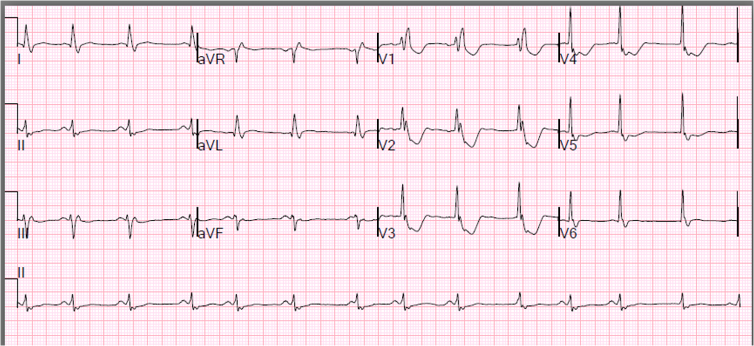
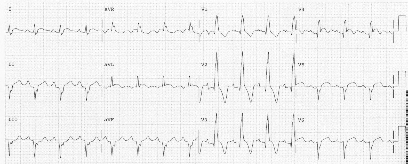
Watch this great presentation.  (For those of you who know my posts and research, you will find some slight differences in Dr. Wang’s approach.)

ST Elevation in Conditions other than Acute MI from HQMedEd on Vimeo.

K. Wang, MD
Clinical Professor of Medicine
Cardiology Division
University of Minnesota

med.umn.edu/cardiology/faculty/wang/home.html

Previous Article

K. Wang Video: The Rhythm Strip Tells the Whole Story

Next Article

Severe Atypical Chest Pain in a Young Woman: Series of Pericarditis ECGs

View Comments (4)
  1. Nice presentation !!! Though im a bit disappointed that Dr K Wang didn't show head to head Benign Repol vs Pericarditis..As i have read, PR depression is not sensitive to pericarditis..The fishhook sign is also not that sensitive for early repol…Moreover, the fishhook is actually the J point which may also present in pericarditis

    Any pearls on how to differentiate these two?

  2. Steve Smith

    Ryan,

    here is K.'s answer: In my experience the “fishhook” is essential to make the Dx of early repolarization. I have seen very minute (a baby) “fishhook” in pericarditis but very rarely. Near the end of my session, one frame showed early repol and the next frame which had composite of 7 cases of ST elevation had pericarditis. The PR segment depression in pericarditis reflects atrial counterpart of ST elevation, i.e. subepicardial injury pattern of the atrial wall and is almost always there.
    Thanks for the comments.
    K. Wang.

    My answer would also include that pericarditis is very unusual. It also is not important to diagnosis it. If you suspect it, do a bedside ultrasound to rule out effusion and look for good pump function, and maybe checkk troponin to look for myocarditis. The main problem with pericarditis is that it can mimic STEMI, but even this is not a big problem because it is so rare. I always say: "you diagnose pericarditis at your peril." because it is more likely STEMI than pericarditis. If not STEMI, the who cares if it is pericarditis or early repol – both are benign.

  3. Wonderfull presentation!
    Is there a way to distinguish right ventricle overload/dilation/ischaemia from acute anteroseptal STEMI based on morphology of V1-V3?

  4. Steve Smith

    Difficult to answer: the morphology is very recognizable subjectively, but hard to give an exact description: anteroseptal MI has slight ST elevation and upward sloping, then sharp downturn of the T-wave, whereas RV overload has a more gradual curve to the T-wave inversion. Also, the best objective way to distinguish them is the presence or absence of T-wave inversion in lead III (presence associated with RV strain).

Leave a Comment

Your email address will not be published. Required fields are marked *