Dr. Smith's ECG Blog

Instructive ECGs in Emergency Medicine Clinical Content

Associate Editors:
— Pendell Meyers & Ken Grauer (2018)
— Jesse McLaren & Emre Aslanger (2022)
— Willy Frick (2024)

editors

PVC or Aberrant Conduction? (Another Guest Post from Dr. Wang)!

Question:
This tracing shows (choose one from the list below)

              
a) atrial bigeminy with aberrant conduction

              
b) ventricular bigeminy
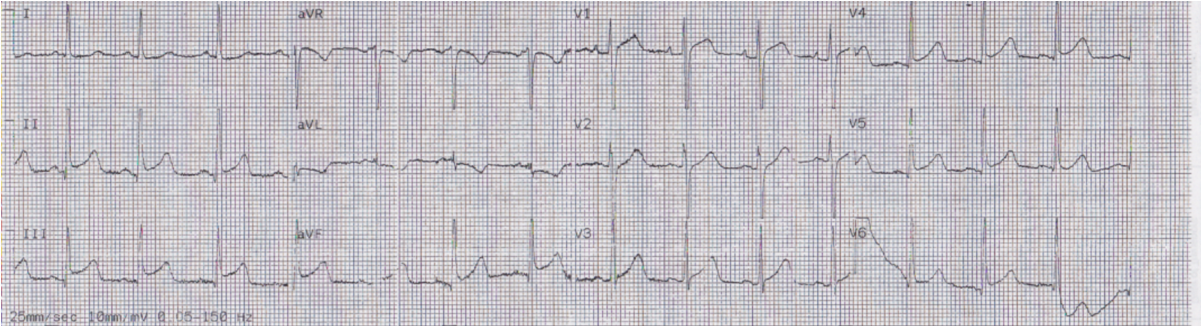
 Annotated ECG:

Answer:  
a) atrial bigeminy with aberrant conduction.

Discussion:
Every other QRS is wide, different looking from the sinus beats and, at a quick
glance, the tracing seems to show ventricular bigeminy. However, careful
observation reveals a prematurely occuring P wave (↓) in front of these wide QRSs and this
is an examle of atrial bigeminy with aberrant conduction. Why does aberrant
conduction ever occur? It is simply because the two bundle branches
(BBs) have different length of refractory periods. If an impulse
occurs when one BB is still refractory while the other has recovered from the
refractory period, the impulse will conduct thru the recovered BB, bypasssing
the refractory BB: aberrant conduction results. Of course, if an impulse occurs
at a time when both BBs have recovered from the refractory period, it will
conduct normally, while if both BBs are refractory, it won’t conduct at
all (non-conducted PAC). This was well diagrammed and explained in the
Medscape ECG of the week posted on 3/7/2012.

Previous Article

Series of Prehospital ECGs Showing Reperfusion

Next Article

24 yo woman with chest pain the morning after binge drinking: Is it Pericarditis?

View Comments (15)
  1. Simon Mortensen

    Nice post again! It seems there's a slight delay through the AV node as well? Is one of the two ventricles generally more likely to have a longer refractory period than the other? We see both LBBB and RBBB patterns in aberrancy, but what's most common?

    1. Steve Smith

      K. Wang answers that: The right bundle branch more frequently has a longer refractory period than the left, and thus aberrant conduction more often manifests as a RBBB pattern.

    2. RBBB is common type of abberency..LBBB is rare.basically all this is type 3 abberrant conduction.Normal finding.Type 4 is always abnormal

  2. If you look at the ST segment in lead lead III it looks like there a small inverted T wave that is followed by a small positive upstroke. I have seen these small positive upstrokes before but have not called them dropped complexes, just assumed that they were part of the repolarization cycle since they were part of the ST segment.
    Anyway, this really helps out because I have been at a loss as to what to call these "PVCs" with P waves. I thought they were some kind of fusion complexes.

  3. But there is qR in V1 and also R>R´ in V1+2 (mostly V2) as well as R/S < 1 in V6 during the "extra"-beats, which if it was about VT vs. SVT with aberrancy would all suggest ventricular origin. How can this not hold true?
    Thanks – S.

    1. Steve Smith

      These are all consistent with RBBB with old MI present (Q-waves). R/S is often < 1 in V6 in RBBB.

      Steve Smith

    1. Because the aberrancy is of BOTH the right bundle and the left anterior fascicle. With left anterior fascicular block, you get left axis deviation. OK?

  4. Is this a true life example? If so, I am so curious. What was the pts. history, and how did they present? Love this by the way! Good for the brain.

    1. Nicole,
      I am sorry to say that the patient was seen years ago and the clinical history is unavailable.
      Steve

  5. structures32

    Hello, Dr. Smith! REgarding this statement on the discussion: "…while if both BBs are refractory, it won't conduct at all (non-conducted PAC)."

    My question is: If I saw a non-conducted PAC on an ECG, I might interpret it as a second-degree AVB type II. How can I differentiate a 2nd Degree AVB from a non-conducted PAC? Thank you very much for your expertise.

    1. You would have to judge as to whether the PAC came early enough to be during the refractory period.

    1. ECG Interpretation

      The reason why some PVCs manifest a "complete compensatory pause" — is that when a PVC does not conduct retrograde — then the next P wave occurs on-time (but is hidden within the PVC) — and the sinus P wave AFTER THAT is also on time — and that's the reason the interval between the 2 sinus beats is twice the normal sinus rate. However, up to half of all PVCs DO conduct retrograde (back to the atria) — in which case the SA node will be reset, and the interval containing the PVC will no longer be "completely compensatory". What happens with PACs — is that because they arise from another site in the atria, they depolarize the rest of the atria (including the SA node) — and this "resets" the cycle. Usually the interval containing the PAC will turn out NOT to be exactly twice the normal R-R interval — but sometimes BY CHANCE the amount of time it takes for the PAC to depolarize the rest of the atria and then for the SA node to fire again will turn out (by chance) to be precisely twice the normal R-R interval — in which case it may seem like there is a compensatory pause with a PAC, when actually this happened by chance … As should be evident — there are LOTS of possibilities — and it's hard to explain with words alone. Hope that helps! — 🙂

Leave a Comment

Your email address will not be published. Required fields are marked *