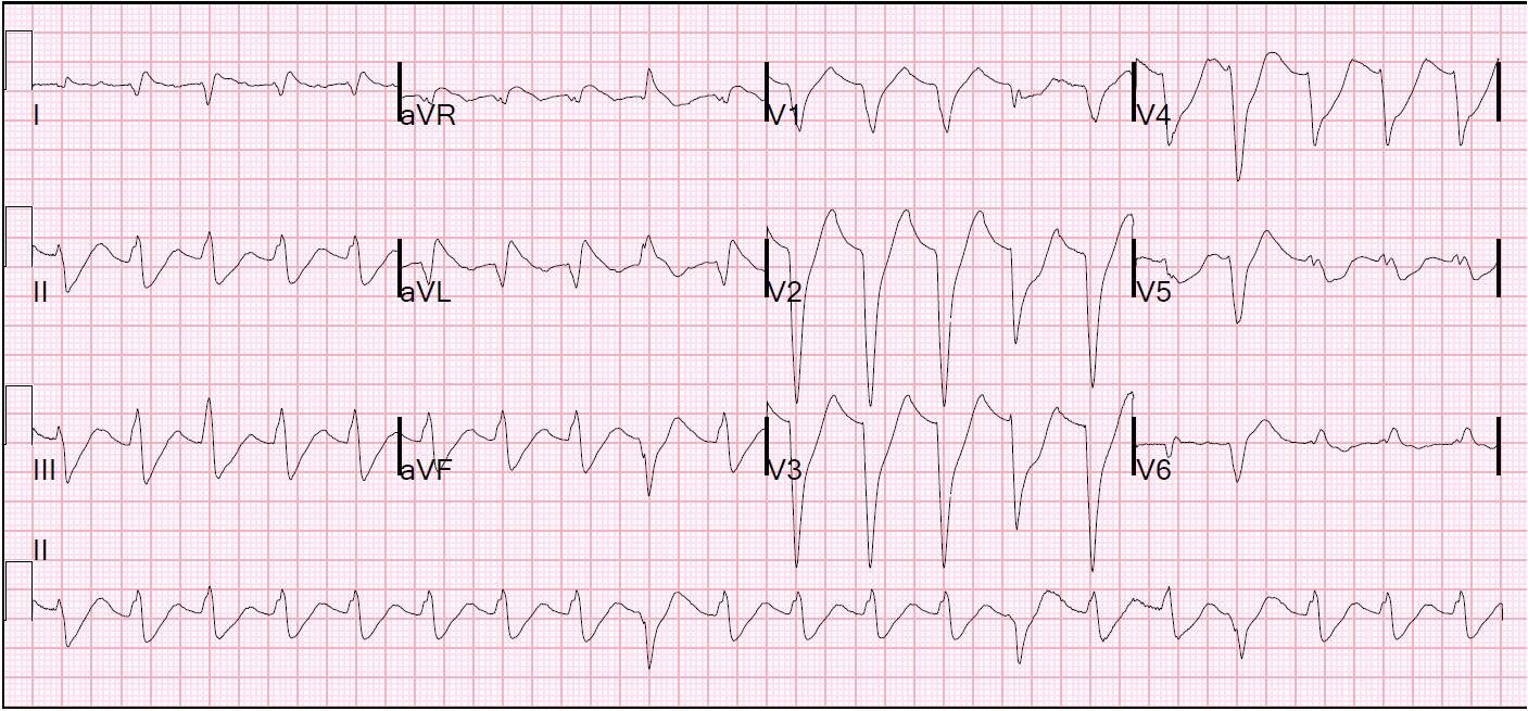
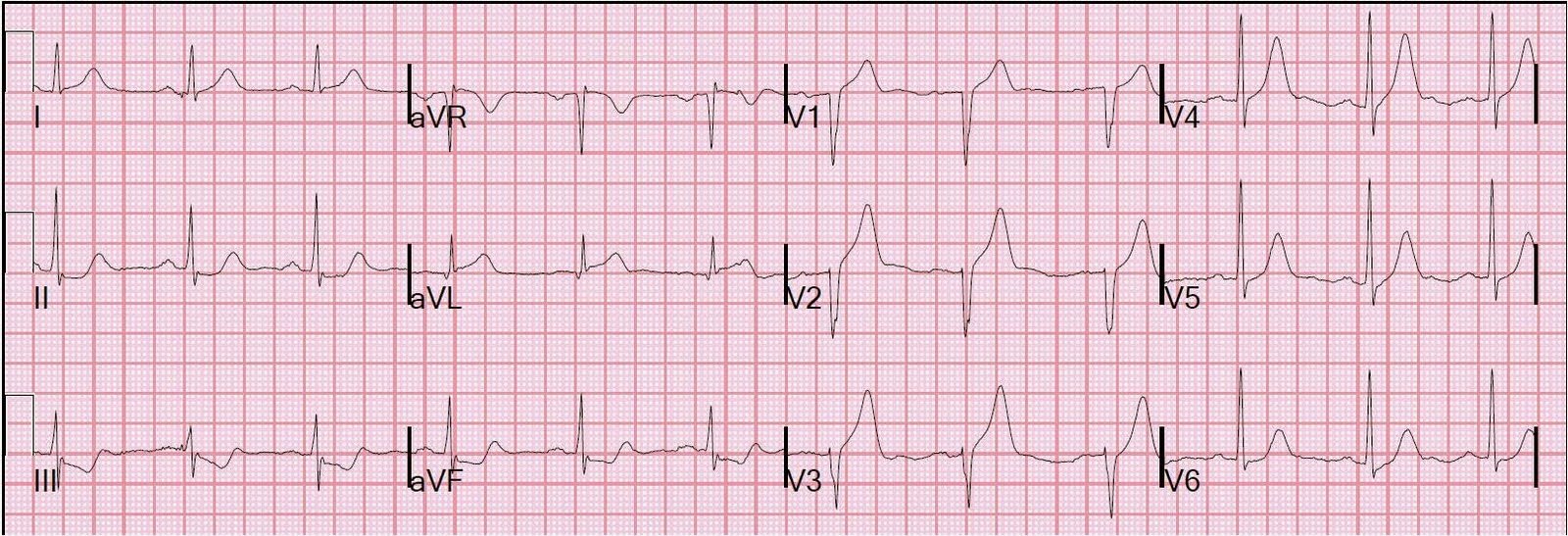
Dr. Smith's ECG Blog

Instructive ECGs in Emergency Medicine Clinical Content

Associate Editors:
— Pendell Meyers & Ken Grauer (2018)
— Jesse McLaren & Emre Aslanger (2022)
— Willy Frick (2024)

editors

RV MI diagnosed by ST elevation in V1

Previous Article

The Importance of Serial ECGs

Next Article

Left Ventricular Aneurysm or Anterior Acute STEMI?

View Comments (7)
  1. Dr. Smith,
    thank you as always for the fantastic education!
    in this case, where the thrombus embolized from proximal to distal RCA, would it be reasonable to say that inverted reperfusion T waves in V1 and V2 may have developed to replace the STE when the proximal occlusion was essentially spontaneously cleared by virtue of the thrombus traveling distally?

    thanks,
    David

  2. ECG Interpretation

    Touche for Dr. Smith – excellent insight provided by this tracing to the site of occlusion in the RCA (that was not initially picked up by the interventionalist . . . ). GREAT case! – : ) Ken Grauer, MD

  3. hi Steve…

    i can just imagine the conversation in the cath lab:
    "dr Heart, its doctor Smith from the ED , again"
    pause, and tremors.
    "damn!, this wasn't even his patient. tell him i'm in the john".
    "dr heart, he knows you're doing the cath now".
    "ok, ok."
    he takes the phone. "Steve! how are you today?"
    the rest is history. very nice case, Steve.

  4. Why there is no sign of Posterior MI?? isnt some STd common in anterior leads; inferior STEMI with RV infarct is it common(without posterior MI)???
    Thank you for the case…

    1. Posterior MI occurs in inferior MI when there are posterior branches of a dominant RCA or dominant circ. RV MI occurs when RCA is occluded very proximally, before the RV marginal branch. It is only associated with posterior MI when there are downstream posterior branches, which occurs maybe 35% of the time.

Leave a Comment

Your email address will not be published. Required fields are marked *