
A 60-something had been having chest “soreness” on and off for one month when she presented with sudden chest discomfort and dyspnea starting about an hour prior to arrival.
Here is the triage ECG.
It was texted to me along with concern for inferior ST Elevation and STE in V1, possible inferior and right ventricular OMI:
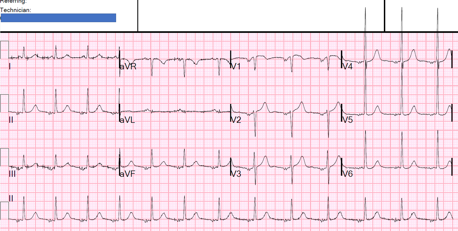
There is a negative P-wave in lead II.
You’ll notice that the P-wave is abnormal everywhere.
This is an ectopic atrial rhythm, and it is low in the atrium such that the atrium is depolarized AWAY from lead II and is inverted.
In any ECG, there might be an atrial repolarization wave, or not. When there is sinus rhythm, with conduction towards lead II, the atrial repolarization wave is negative, resulting in PR depression and depression of the first part of the ST segment (see schematics below). This is why we measure the ST segment relative to the QRS onset (PQ junction): both intervals may be depressed by a normal atrial repolarization wave.
In pericarditis, the atrial repolarization wave is exaggerated; hence, we get excessive PR depression.
In low atrial rhythms, on the other hand, when the atrial impulse is traveling from inferior to superior, the atrial repolarization wave is positive, and can result in a mimic of ST Elevation.
This is what is happening in inferior leads, but ALSO in V1.
Ken Grauer says this is called the “Emery Phenomenon,” but I can’t find that in any search.
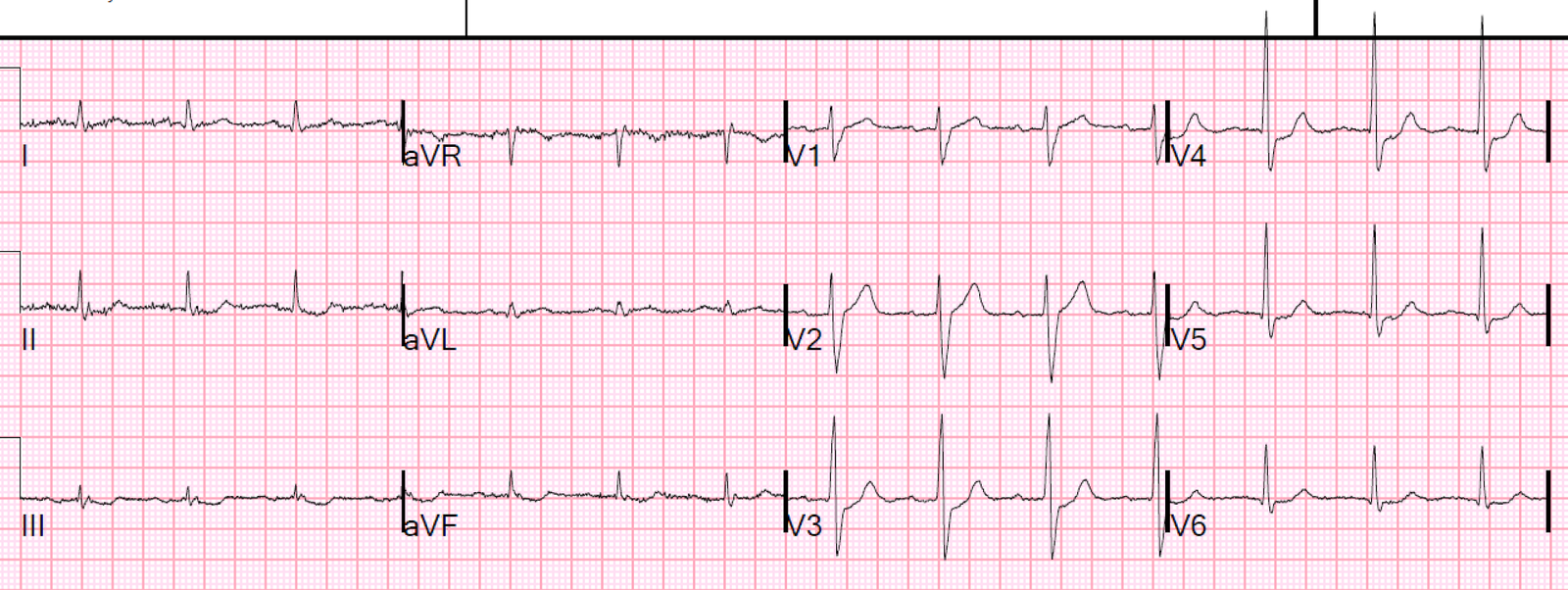
Here is a magnification of V1:

Here it is with the atrial repolarization wave outlined by a circle:

Case continued:
I responded that this was unlikely to be OMI, that it was probably an atrial repolarization wave, and to get serial EKGs and troponins.
The patient ruled out for MI, and probably had chest wall pain.
See below for more Learning about the Atrial Repolarization wave.
Here are some schematics:

Normal atrial repolarization wave
This schematic is from my book, The ECG in Acute MI

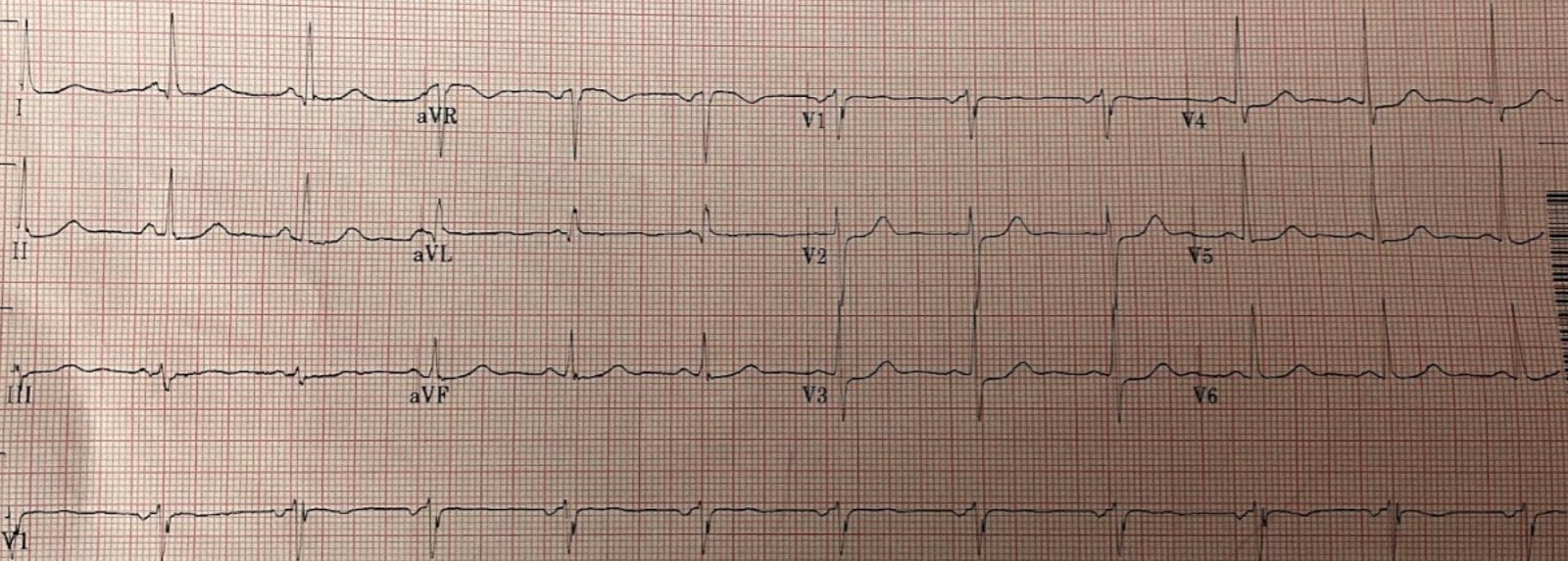
Here is another recent case:
A man in his sixties with chest pain

At this LINK, there is an extensive discussion by Ken Grauer
Here you can see how the atrial repolarization wave can mimic what appears to be significant ST Depression:
Look at this ST Depression (It’s not real)

this is terrible, Steve. Just when i was starting to become slightly "comfortable" reading ecg's, and posterior wall MIs in particular, you show me these cases , and that last ecg in particular. i definitely, (and now i learn , incorrectly) would have fought for a stat cath lab activation on that last ecg. (a PWMI that isn't).
i need to study more.
thank you , again.
excellent blog.