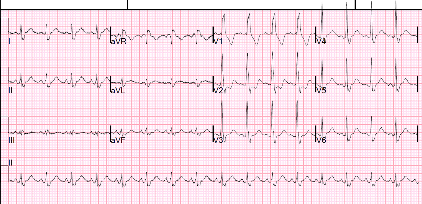
I don’t have the clinical presentation, but the ECG is diagnostic on its own:
 |
| See question and answer below |
What does this tracing show? Choose
one from the list below.
a) Hyperkalemia
b) Hyperkalemia and
hypocalcemia
c) Hyperacute ischemic
changes
d) Normal varient
Answer: b) Hyperkalemia and
hypocalcemia
Discussion: T waves are tall, tented
and pointed, indicating hyperkalemia. That T waves are “pushed out”
by a long ST segment especially noticeable in V5&6, characteristic of
hypocalcemia, too. The serum K was 6.6 mEq and the serum Ca was 5.3 mg/dL in a
patient with chronic renal failure, which is known to cause these combination
of electrolyte problems. It is not hyperacute ischemic changes because the tall
T waves are tented (the base of the T wave becomes narrow and pointed).
There is some asymmetry of the T-waves, especially in V3 and Lead II, suggestive of normal variant, but the T-waves are far too “tented” and narrow-based for simple normal variant. The
differential Dx of tall T waves are well outlined in the book “Atlas of
Electrocardiography” (amazon.com books), page 171.