A middle-aged male presented with acute onset of typical ischemic type chest pain.
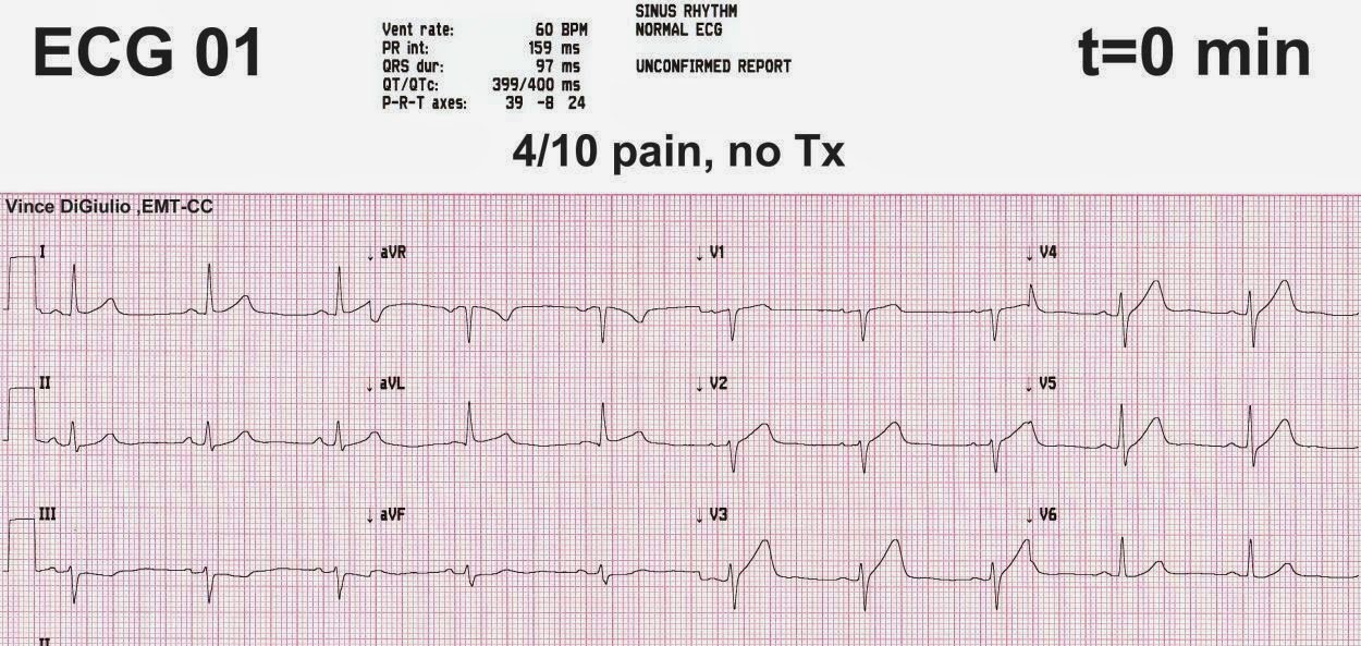
Here is his initial ECG:
 |
| What do you see? |
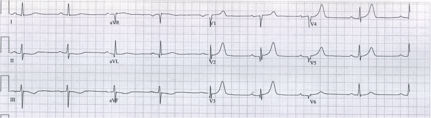
He had a previous ECG from 10 years prior:
 |
| This shows either subaute anterior MI (Q-waves, ST Elevation, T-wave inversion) or acute MI superimposed on previous MI. LV aneurysm is possible but unlikely (no QS-waves, significant T-wave amplitude, even though largely negative). |
So we know he has LAD disease and previous MI.
The acute ECG (first one at top) clearly shows the previous MI (Q-waves). The T-waves are upright. Is this pseudonormalization? No. Over weeks to months and certainly years, inverted T-waves of MI normalize as part of the natural history of the ECG in MI (Although, in LV aneurysm, they often remain with shallow invertion). So before this acute event the T-waves were likely upright.
But these previous T-waves were not upright and hyperacute, as they are here in the top ECG: the T-waves in that first ECG are suspiciously large, and there is some hint of reciprocal ST depression in inferior leads. These findings need aggressive investigation with frequent serial ECGs or immediate echocardiography. They could represent LAD occlusion.
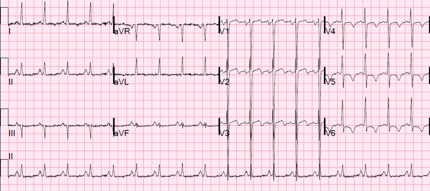
The pain continued and another ECG was recorded 1 hour later:
 |
| What is the diagnosis? |
These are now subtle de Winter’s hyperacute T-waves; see the subtle ST depression in V3.
These were not seen or appreciated. The initial troponin returned at less than 0.04 ng/mL. The patient was admitted to the hospital without angiography in spite of continued chest pain.
A second troponin at 6 hours returned at 0.7 ng/mL (elevated). He was diagnosed with NonSTEMI. The pain continued.
A third ECG was recorded at 7 hours:
 |
| Now the T-waves are less acute, but there are deepening Q-waves and loss of R-wave height |
The troponin I was 16 ng/mL at hour 13.
Here is the 13 hour ECG:
 |
| T-waves are smaller yet, especially in V4 |
He went for cath and it showed 100% ostial LAD occlusion. There was no mention of collateral circulation.
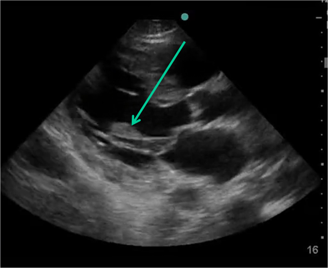
Echo: Anterior wall motion abnormality with EF of 35-40%, although it is unknown what the EF was before this event. There was no myocardial wall thinning to suggest that this WMA was due to old MI, though the previous ECG is fairly conclusive about that.
Here is a post cath ECG at 26 hours:
 |
| Now there is also some inferior ST elevation with reciprocal ST depression in aVL. This is highly suggestive of inferior MI. The RCA was anomolous, arising from the left coronary cusp, and it is possible (though not mentioned) that it was affected during the procedure. There is no more information on this. |
Diagnosis: LAD occlusion in the context of previous MI, and manifesting subtle de Winter’s T-waves.
Here are some more examples of the wide variety of de Winter’s T-waves from LAD occlusion: