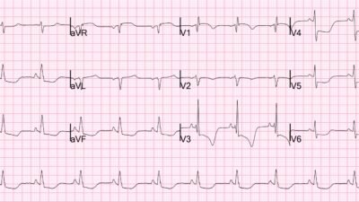
A 50 yo male had out of hospital cardiac arrest. He was put on rapid Mobile ECMO (ECPR) due to absence of initial ROSC. It is unknown whether dual sequential defibrillation was used prior to ECPR.
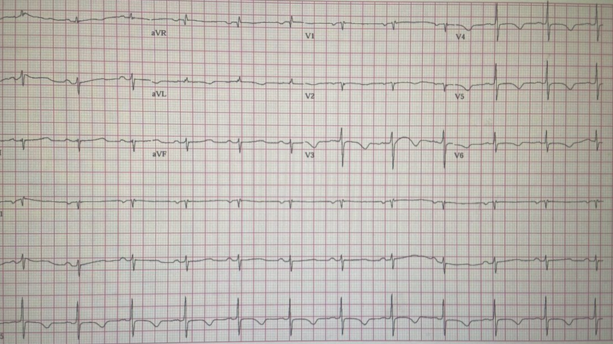
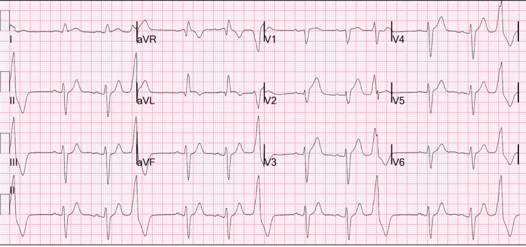
Later, he had ROSC and had this ECG:
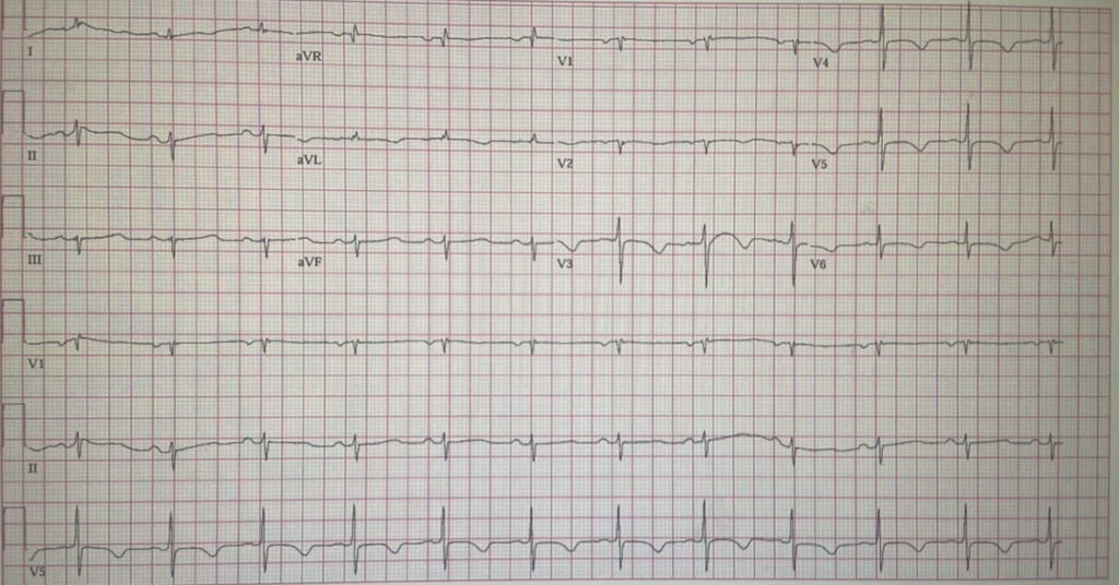
— What do you think? —
= = =
Here is the PMcardio Queen of Hearts AI ECG diagnosis:
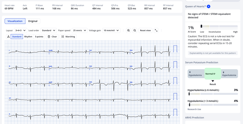
She sees neither active nor reperfused OMI, nor subendocardial ischemia. And there certainly is not a STEMI (OMI that meets STEMI criteria)
I also did not see any evidence of active OMI, though it certainly could be a reperfused OMI.
At the senders institution, all patients undergo Coronary angiogram if they are cannulated for VF arrest.
Angiogram showed 100% Circumflex occlusion (TIMI-0). It was opened and stented.
The patient survived.
Learning Points:
- Approximately 10% of active OMI do not have an ECG that is diagnostic of Active OMI. One must always keep one’s index of suspicion high.
- Due to the fatally flawed COACT and TOMAHAWK trials, guidelines discourage cath lab activation in the absence of NSTEMI high risk features. Even if a patient has ECG evidence of active OMI (which this one does not), if it does not meet “STEMI” criteria, then the patient should not undergo angiography.
- The guidelines do actually make an exception for patients on ECMO, but that is in the fine print.
See this post for critique of COACT and TOMAHAWK: Guidelines would (erroneously) say that this patient who was defibrillated and resuscitated does not need emergent angiography.
I’ll show the ECG here:
Some notes on ECPR for cardiac arrest
ECPR (AV ECMO) is controversial for cardiac arrest. One objection to all the studies is that they did not compare with dual sequential defibrillation, only with standard defibrillation. That includes the most recent study, just published this month (see below). It certainly can only be done in select institutions with extensive training and it is very expensive.
Prehospital ECMO, from Nov 7, 2026 (full text link):
Prehospital Extracorporeal Life Support in Refractory Out-of-Hospital Cardiac Arrest
This is a summary from Open Evidence:
ECPR (VA-ECMO during ongoing CPR) for refractory VF is an established but selective rescue therapy. Guidelines say it is reasonable for selected patients in experienced systems, not as routine care. The AHA gives it a Class 2a recommendation for select patients whose arrest has a potentially reversible cause. [1] Refractory VF/VT out-of-hospital cardiac arrest (OHCA) is the population with the strongest evidence, but the randomized trials disagree. [1-2] Prehospital (on-scene) cannulation is still investigational. Current practice relies mainly on rapid field transport to an ED or cath-lab ECMO team.
Randomized evidence (all OHCA)
- ARREST (single centre, Minnesota, refractory VF/VT): refractory meant VF/VT persisting after 3 shocks and amiodarone 300 mg, with transport time under 30 minutes. Patients went straight to the cath lab for ECMO and coronary angiography/PCI. Survival to discharge was 43% vs 7%, and the trial was stopped early for superiority. The authors note it depended on a high-volume centre and an EMS system restructured so the ECMO team could deploy within about 20 minutes of the 911 call. [1-3]
- Prague OHCA (single centre, 264 patients, any rhythm; about 40% non-shockable): used intra-arrest transport, ECPR and invasive assessment. Favorable neurologic outcome at 180 days was 32% vs 22%, which was not significant, and the trial was stopped for futility. In a secondary analysis, patients with an initial VF rhythm and those without field ROSC after prolonged CPR did benefit. [1-2]
- INCEPTION (10 Dutch centres with variable ECPR experience, initial ventricular arrhythmia): favorable neurologic survival at 30 days was 20% vs 16% (OR 1.4, 95% CI 0.5–3.5). Time to ECMO was longer and cannulation failed more often than in the single-centre trials. [1-2]
- Pooled RCT data: short-term favorable neurologic outcome improved significantly. Mid-term neurologic outcome and survival showed only non-significant trends, and major bleeding increased. [4]
= = =
======================================
MY Comment, by KEN GRAUER, MD (11/21/2026 ):
The patient in today’s case was successfully resuscitated from OHCA (Out-of-Hospital Cardiac Arrest). The patient was taken to a facility that performs cardiac cath on all survivors of OHCA. The cath showed 100% Circumflex occlusion (TIMI-0) — which was successfully stented, and the patient survived.
- The above said — it is not standard practice in all U.S. institutions to promptly take all survivors of OHCA to the cath lab. The reason is simple: Doing so is not benign. To many, the potential fate of hypoxic encephalopathy (futility of care) is worse than death.
- An additional consideration arises in assessing which OHCA patients with successful ROSC (Return Of Spontaneous Circulation), but with uncertain neurologic status — should qualify for prompt cath. This is because many of these OHCA survivors are resuscitated at institutions without ready access to 24/7/365 cath service. This complicates decision-making with time-consuming transport issues when trying to determine which OHCA survivors have at least reasonable potential for positive outcome.
= = =
Which Patients with OHCA should go to Cath?
The answer to the question of which patients with ROSC following OHCA should go to the cath lab is challenging. There is no universal agreement. I found these articles to be informative:
- Tamis-Holland et al (Circulation 149(5), e274-295, 2024).
- Staer-Jensen et al (Circ: Cardiovasc Intervent 8(10), 2015).
= = =
First — some Statistics:
- OHCA is a leading cause of death, accounting for ≈50% of all cardiovascular events. The overall prognosis of victims of OHCA is poor — with less than 10% surviving to hospital discharge.
- The “better news” — is that survival from OHCA may be increased to ~30%, if common sense favorable parameters are associated with the episode. Survival with a positive neurologic outcome is highest among patients with OHCA who: i) Present with a witnessed shockable rhythm; — ii) Receive prompt bystander CPR; — iii) Achieve ROSC within 15 minutes of arrest — and, iv) Show an acute STEMI on their initial ECG done following ROSC.
- There is agreement that survivors of OHCA who promptly regain consciousness — and whose initial ECG shows an acute STEMI should go to the cath lab. But in the absence of these favorable predictors — consensus is lacking. And there are caveats …
= = =
Caveats in the “Selection” Process …
- The initial ECG following ROSC is not reliable for detecting all post-arrest patients who potentially benefit from prompt reperfusion. This is because ECG findings of acute ischemia are not always immediately seen — and there are false-positive ECGs …
- This highlights the importance of the History (if any history is known at the time the patient is evaluated).
- Does the patient have known coronary disease?
- Was the arrest witnessed?
- Was CPR promptly started?
- Did the patient have chest pain before collapse?
- In patients without frank ST elevation on their initial post-resuscitation ECG — the literature reports a wide range (3%–58%) among those who demonstrate cath findings consistent with acute coronary occlusion.
- ECGs obtained immediately after ROSC lack specificity. The time from arrest, and the time from when ROSC is achieved will significantly affect the reliability of the 1st post-ROSC ECG for detecting which patients had acute coronary occlusion as the cause of their arrest (ie, The number of false-positive STEMI diagnoses progressively decreases the longer one waits before recording the 1st post-resuscitation ECG — with the optimal time reflecting a balance between not wanting to wait too long vs not wanting too many false-positive tracings).
- Observational data suggests waiting ~8 minutes after achieving ROSC may strike that optimal balance between minimizing false-positive STEMI ECGs (caused by poor tissue perfusion from transient low flow) — but not waiting “too long” before cath with PCI in survivors whose OHCA was indeed caused by acute coronary occlusion (Baldi et al — JAMA NetwOpen 4(1):e2032875, 2021) — (2025 AHA Guidelines for Cardiopulmonary Resuscitation) — (Savastano et al — Resuscitation 192, 109971, 2023).
- Alternatively — One might choose to obtain the post-ROSC ECG as soon as this is feasible, and to repeat the ECG within a few minutes if the initial tracing showed ST elevation (so as to minimize false-positive results) — vs the approach selected in today’s case of performing cardiac cath for all survivors of OHCA.
- False-positive STEMI tracings are especially common in patients with hyperkalemia and/or mental impairment (which of course are common accompaniments in cardiac arrest).
- Initial neurologic assessment post-ROSC is notoriously inaccurate for predicting ultimate neurologic outcome in OHCA survivors (ie, Some patients ultimately recover normal or near normal neurologic function despite an initial period of unresponsiveness with seemingly terminal neurologic predictors including myoclonus and absent pupillary reflexes).
- As a result — Neurologic assessment should not be used as the sole criterion for early decisions regarding cardiac catheterization.
- That said — awareness of those clinical features associated with poor neurologic outcome may help when considering the overall decision-making process (ie, These adverse clinical features include advanced age, unwitnessed arrest, absence of bystander CPR, nonshockable rhythm on initial assessment, and prolonged duration of cardiac arrest before ROSC).
- Bedside Echo may help to identify OHCA survivors with acute coronary occlusion (ie, if a localized wall motion abnormality is seen) — and Echo may help to identify the ~5% of cardiac arrests triggered by acute PE (ie, if marked RV dilatation is seen). That said — overall sensitivity and specificity of bedside Echo is limited when performed in OHCA survivors.
- NOTE: An important clue to acute PE as the cause of OHCA is a presenting ECG rhythm of PEA or asystole.
- BOTTOM Line: Predicting which victims of OHCA in whom ROSC is achieved will ultimately survive with favorable neurologic function remains an imperfect science. But — timely cath with PCI in those patients who do have acute coronary occlusion as the cause of their arrest (as was present in today’s case) — will clearly save a selected number of patients.
= = =
Application to Today’s ECG …
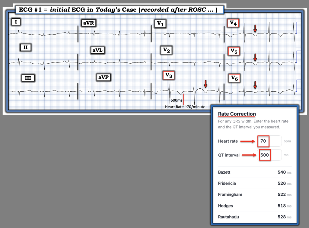
For clarity in Figure-1 — I’ve reproduced the post-ROSC ECG from today’s case. We are not told how much time passed after achievement of ROSC until the recording of this tracing.
- The rhythm in ECG #1 is sinus at ~70/minute.
- The QTc is prolonged (As shown in Figure-1 — Use of our on-line QTc Calculator faciliates rapid determination of the QTc).
- Moderately deep, symmetric T wave inversion is seen in leads V3-thru-V6 (RED arrows) — with more subtle T wave inversion also seen in high-lateral leads I and aVL.
My Thoughts: This post-ROSC tracing illustrates the difficulty inherent in interpreting ECGs in the low-flow state following resuscitation from cardiac arrest.
- The tracing is clearly not normal.
- The T wave inversion seen in multiple leads might certainly reflect reperfusion T waves.
- But — ST elevation is not present …
Bottom Line: Realizing that we do not know how much time passed from establishment of ROSC until the recording of today’s tracing — this case clearly illustrates an example of the benefit of performing timely cardiac cath routinely in OHCA survivors deemed to have potentially salvageable neurologic status.
= = =
Figure-1: Today’s ECG — obtained following ROSC.
= = =
= = =