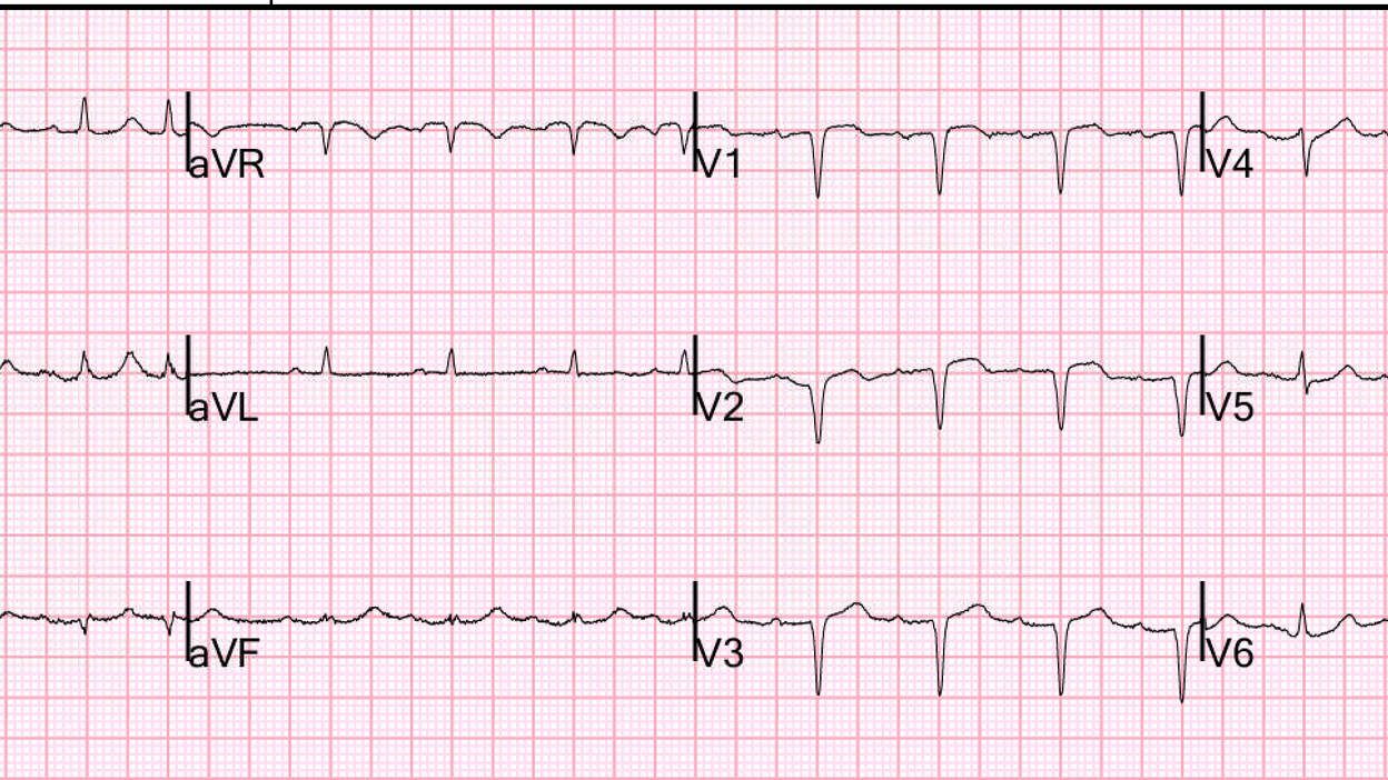
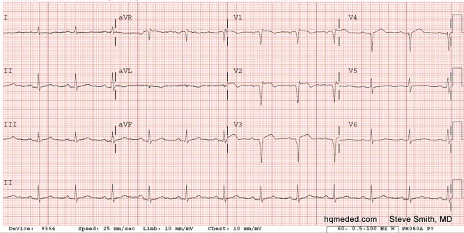
An 80-something presented after 12 hours of constant chest pain which rendered him unable to sleep.
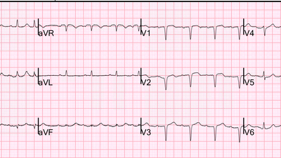
Here is his ECG:
— What do you think? —
= = =
= = =
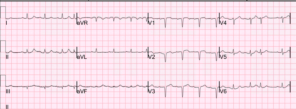
To me (and to the treating physicians) this looks like either: 1) anterior LV aneurysm from completed old anterior transmural infarct (QS-waves with some ST Elevation +/- shallow T-wave inversion); — or — 2) Subacute anterior OMI.
- It is very difficult to tell the difference. In both cases there is completed or nearly complete transmural infarction.
Here is the PMcardio Queen of Hearts AI model:
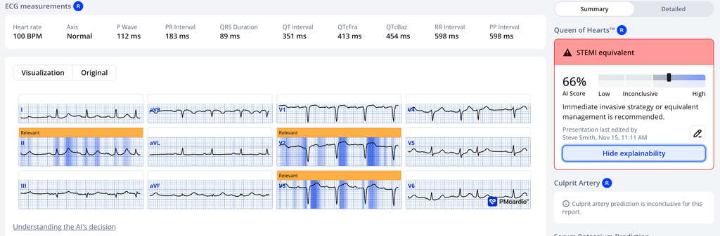
Our newest model (unavailable), which can diagnose LV Aneurysm (“old MI”) still thinks this is acute. She gives it 85% for Active Acute MI and 36% (indeterminate) for Old MI.
The Queen thinks this is acute (subacute), not LV aneurysm.
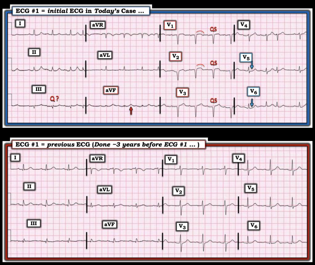
Here is the previous ECG from 3 years prior:
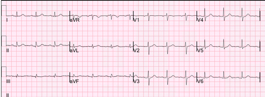
So this MI is clearly new in the last 3 years.
If it is a new MI, then it is subacute with a lot of damage, and an initial troponin should be very high. If it is due to an infarct. Subacute MI is very consistent with 12 hours of constant chest pain.
If it is old MI only, then first trop should be nearly normal, though such an ECG would often be associated with heart failure and a mildly elevated troponin due to chronic myocardial injury
If it is a new MI superimposed on previous large MI, then troponin will not be particularly helpful.
The good thing is that one can wait for the first troponin because a short delay will not result in much more myocardial death.
- The providers did wait for the first troponin.
- This differential diagnosis was resolved with the return of the first troponin at > 60,000 ng/L
- So this is a subacute MI which began when the patient’s chest pain began 12 hours ago.
Cath lab was activated.
- LAD is a medium to large caliber vessel with 95% stenosis in the proximal segment and TIMI II flow beyond.
Echo:
The estimated left ventricular ejection fraction is 35-40%. Normal left ventricular cavity size and wall thickness. Regional wall motion abnormality: akinesis apical segments, hypokinesis mid inferoseptum, hypokinesis mid inferior, mid anterior, midanteroseptum.
Other examples:
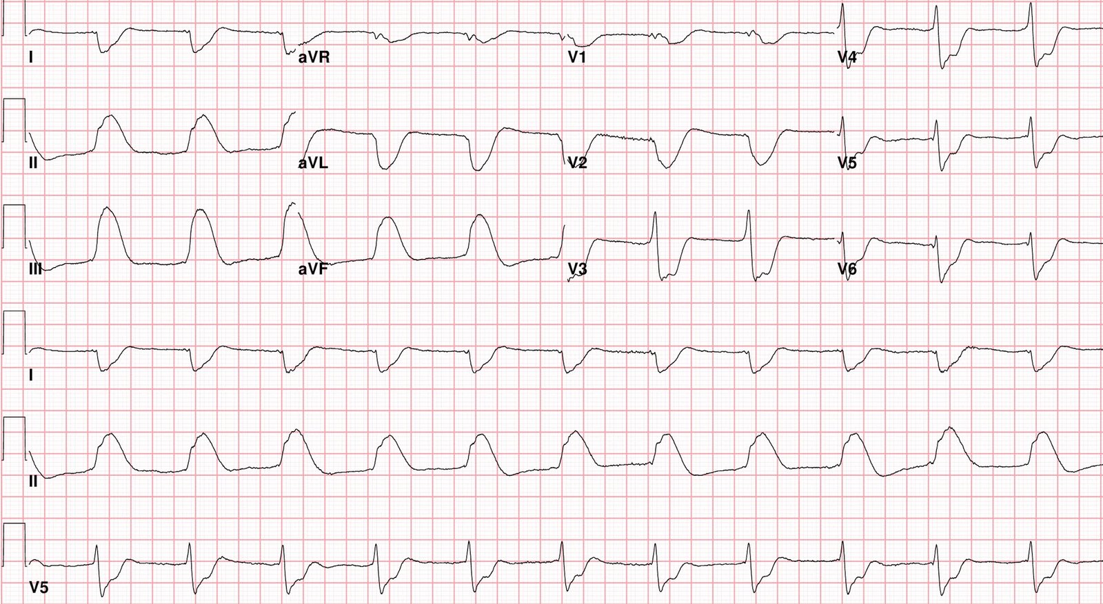
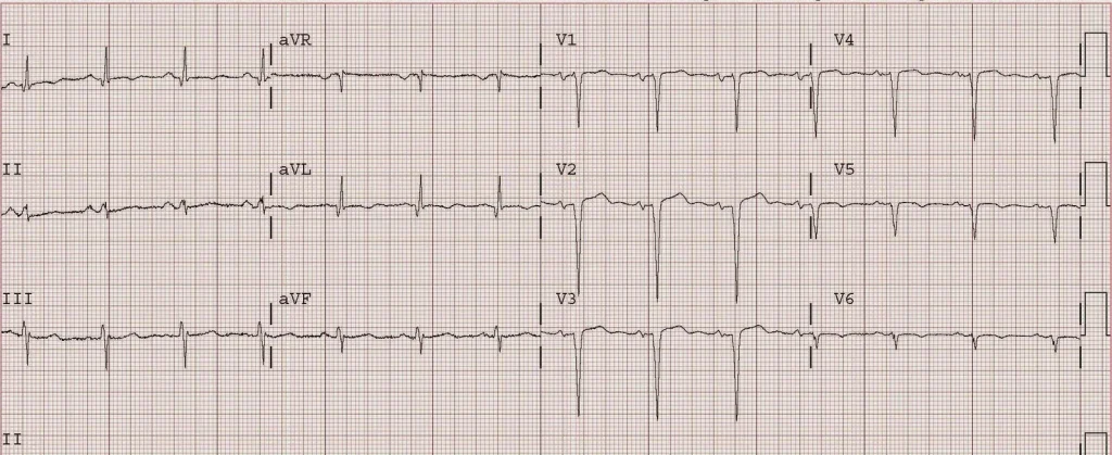
Classic LV aneurysm (persistent ST elevation after previous MI)
Case 1. Classic anterior LV aneurysm morphology (Persistent ST elevation after previous MI).
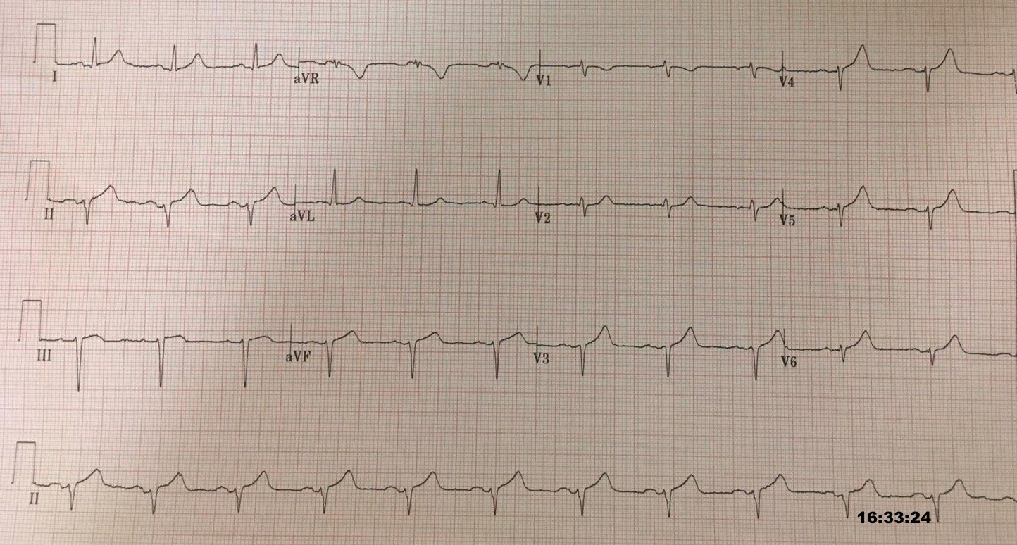
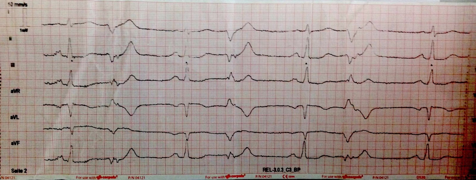
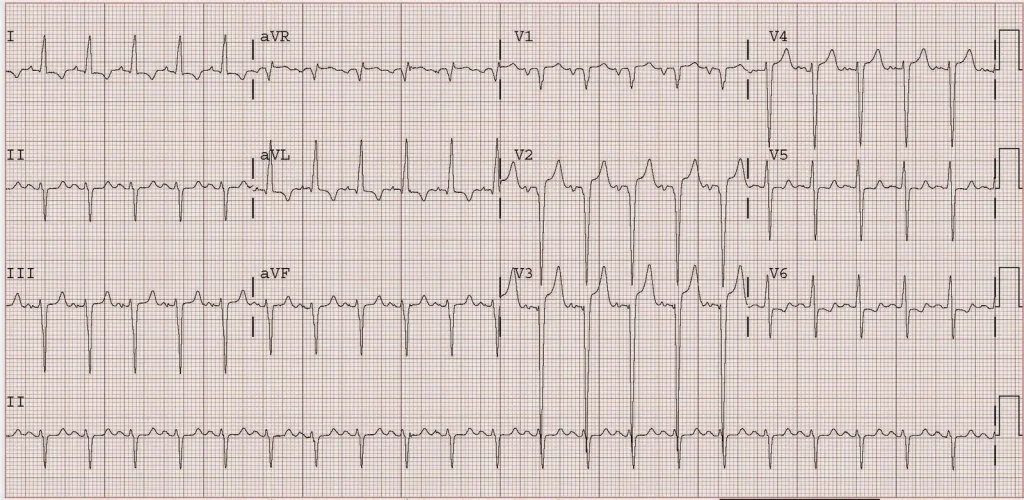
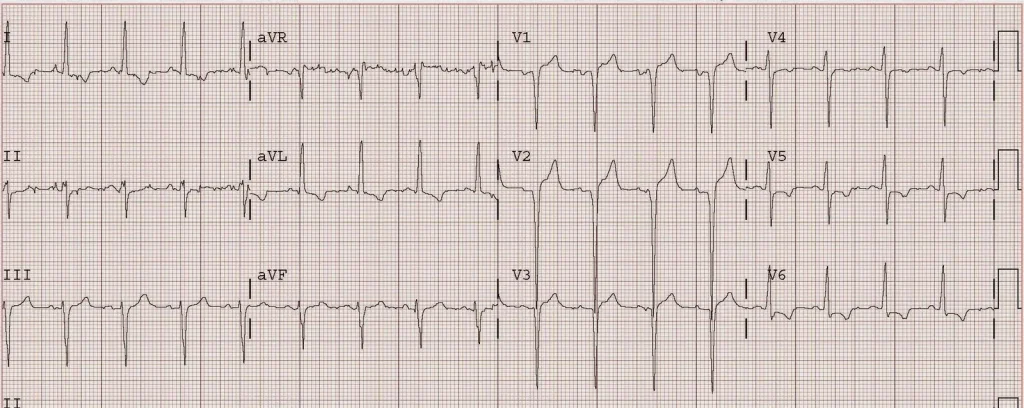
Case 2. Exaggeration of persistent STE after old MI by tachycardia
With tachycardia:
After tachycardia managed:
= = =
======================================
MY Comment, by KEN GRAUER, MD (11/16/2026 ):
Today’s case raises a question that periodically surfaces regarding whether a patient’s initial ECG is the result of: i) LV aneurysm from previous anteroseptal infarction; — or — ii) Recent LAD OMI. As per Dr. Smith — it’s often difficult to distinguish between these 2 entities (See the September 11, 2024 post, among others).
- For clarity in Figure-1 — I’ve reproduced the 2 ECGs in today’s case, highlighting findings of interest in the initial ECG.
= = =
Today’s initial ECG …
The rhythm in ECG #1 is sinus at a rate just under 100/minute. There is one PAC ( = the 3rd beat in simultaneously-recorded leads V4,V5,V6). All intervals (PR-QRS-QTc) and the frontal plane axis are normal. There is no chamber enlargement. Overall QRS amplitude is low (See the August 3, 2025 post — for more on the ECG finding of low voltage). But the findings of most concern include the following:
- QS waves in leads V1,V2,V3 — with no more than a very small r wave in lead V4.
- ST segment coving in leads V1,V2 — with slight ST elevation in lead V2.
- Subtle suggestion of ST depression in leads V5,V6.
- A potentially hyperacute T wave in lead aVF (with assessment made difficult by tiny QRST amplitude in this lead — though with a T wave in this lead that looks to be disproportionately increased in size compared to that tiny QRS).
- The question of a large Q wave in lead III (vs the possibility of an initial small positive deflection [r wave] in this lead).
- My Thoughts: The finding of QS complexes in each of the first 3 chest leads, in association with no more than a tiny r wave in lead V4 — suggests there has been anteroseptal infarction at some point in time. While fully acknowledging the difficulty distinguishing between “old” vs “new” vs “new superimposed on old” — I thought the subtle findings in the above noted bullets suggested the need to consider recent OMI until proven otherwise.
As per Dr. Smith — the “Answer” to today’s case was forthcoming with 3 additional pieces of information. These were:
- The History: This 80-something year old man presented to the ED for 12 hours of severe chest pain that kept him from sleeping.
- Access to a Previous ECG: Comparison between serial ECGs is greatly facilitated by the simple step of placing both tracings next to each other. Doing so in Figure-1 confirms that the loss of R wave in leads V1-thru-V4 is new since the previous ECG that was done 3 years earlier.
- The 1st Troponin Result: Elevation to a level > 60,000 ng/L confirmed the need for prompt cath.
= = =
Bottom Line:
It will not always be possible to distinguish between “old MI with LV aneurysm” vs “new” or “recent” OMI vs “new superimposed on old” on the basis of a single ECG. Today’s case illustrates steps that may facilitate making this distinction.
= = =
Figure-1: The 2 ECGs in today’s case.
= = =
= = =