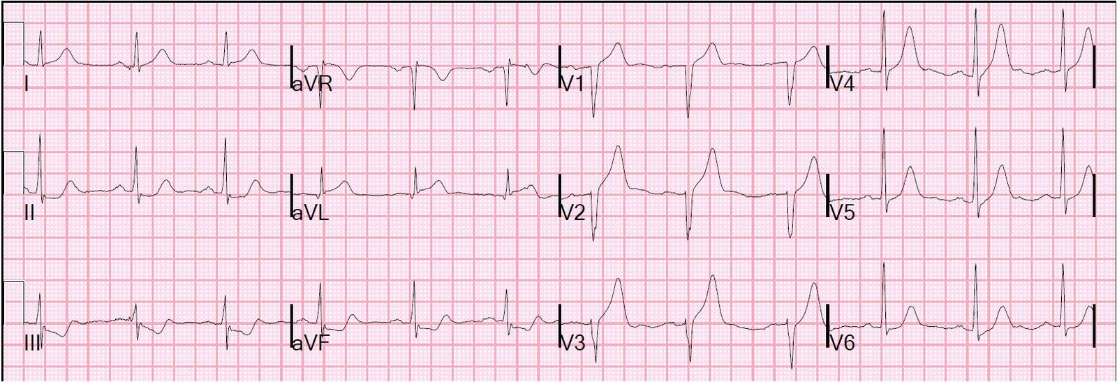
One reader who trained in France learned that a good way to see the ST elevation better is to look at a mirror image of the ECG (upside down). I have done this in order to see that the ST depression in V2 of posterior STEMI is ST elevation when viewed upside down (as it would be recorded from a posterior aspect). I have never seen it done to highlight inferior reciprocal ST depression and to highlight anterior hyperacute T-waves.
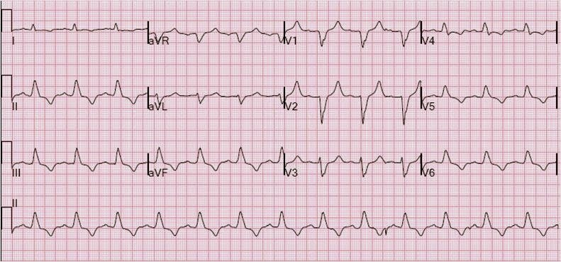
This is a case I posted last week: https://drsmithsecgblog.com/2011/01/hyperacute-t-waves-missed-by-computer.html
 |
| Hyperacute T-waves in V2-V4, see full description at previous blog post |
 |
| Upside Down Mirror Image. Notice now that what was reciprocal ST depression in II, III, aVF is now ST elevation. Also notice that the hyperacute T-waves, when pointing down, look to have ST depression. |