
A middle-aged woman presented with symptoms of mild ischemic stroke. There were no chest symptoms.
An ECG was recorded as part of the workup:
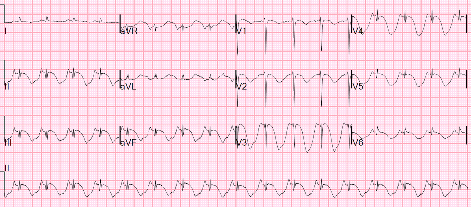 |
| Bizarre inverted T-waves in nearly every lead, with greatly prolonged QT interval. QT = 560 ms. Bazett corrected QT = 727 ms These are classic for CNS catastrophe, especially hemorrhagic stroke They are unusual in a small ischemic stroke. |
This has been called the “spiked helmet sign,” in this case, and in this case.
An Echocardiogram showed apical WMA and both LV and RV thrombi.
The stroke had a very low NIH score and did not receive any reperfusion the therapy for this.
Incredibly, no troponins were ever measured.
A CT Coronary Angiogram showed normal coronary arteries.
She was found to have cancer.
What happened?
There are two at least two possible scenarios:
1. She had stress cardiomyopathy for an unkown reason, developed LV and RV thrombi, which embolized to her brain.
2. She developed LV and RV thrombi due to cancer. These then caused the stroke, which caused the stress cardiomyopathy.
I believe the latter is more likely, as T-waves like this are most commonly seen in CNS catastrophes. A mild stroke is normally not enough to cause this (it is not enough of a CNS catastrophe), but I think this goes to show how intimate the connection between the brain and the heart can be.
3 days later

4th day

Here is a 2020 article on the “Spiked Helmet Sign”
https://www.ahajournals.org/doi/10.1161/CIRCULATIONAHA.120.047427#.XvE165nOf90.twitter

