A woman in her 50’s had a v fib arrest and required defibrillation 10 times before resuscitation to an organized rhythm.
Here was the monitored rhythm:
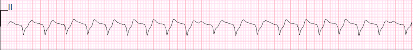 |
| ventricular tachycardia? |
There are two clues that it is not:
1) The rhythm is irregularly irregular, which strongly suggests atrial fibrillation
2) There is a “shelf” on the upslope of the S-wave. This suggests that it is ST segment, not part of the QRS.
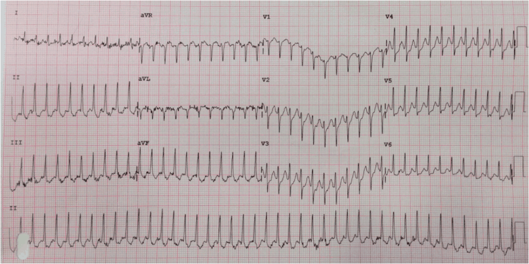
Here is the 12-lead:
 |
| Here you can see in leads V4-V6 that the QRS is indeed narrow. All of the apparent width is really ST segment depression, as well as ST elevation in aVR and V1. |
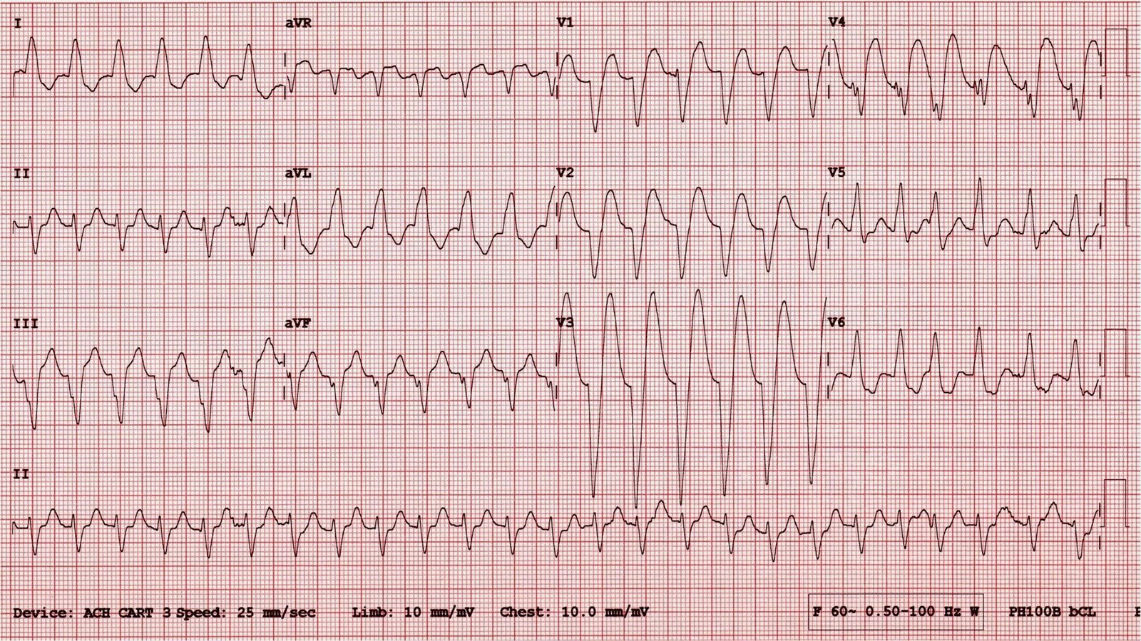
Here I have placed lines to demonstrate the end of the QRS in all leads, using lead II across the bottom for orientation:

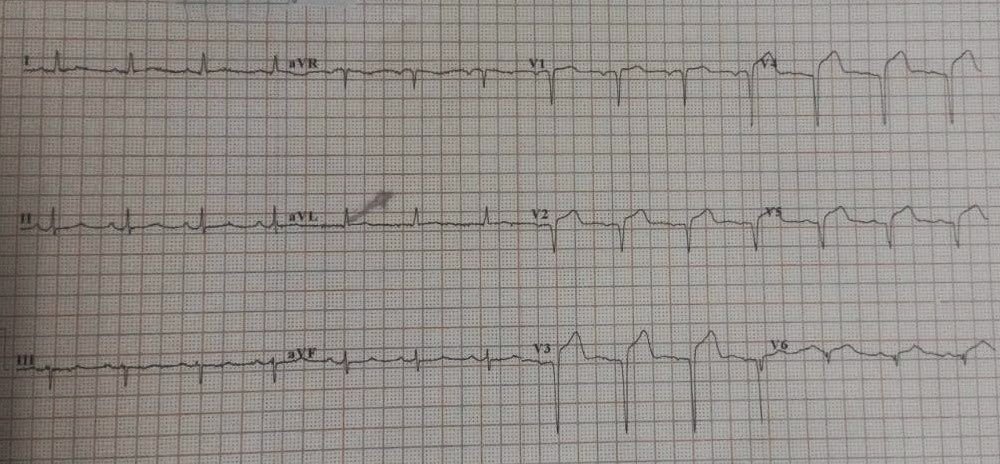
After resuscitation, her ECG normalized:

She was found to have an 80% LAD lesion, with open artery and good flow. This was not definitely a culprit, and not definitely the etiology of arrest.
Lessons:
1. In the presence of ST deviation, the rhythm strip may deceive you into believing there is a wide complex.
2. ST depression is often seen immediately after resuscitation from cardiac arrest. After a short period of stabilization, it will resolve if there is no underlying acute ischemia