Dr. Smith’s ECG Blog Dr. Smith’s ECG Blog
  • Learn
    • Teaching Images
    • Lectures + Podcasts
    • Rules + Equations
    • The OMI Quizzes
  • Tools & Guides
    • OMI Pocket Guide
    • QT Calculator for Wide QRS/LBBB
    • External: Cardiac Cath Guide (Notion)
    • External: OMI AI: The Queen of Hearts
  • Research & Resources
    • OMI Literature Timeline
    • OMI Facts and References
    • Resources
    • Technical Misadventures: Lead Reversals
    • Neuroprotective CPR / Head Up CPR
  • About
    • About
    • The OMI Manifesto

Archives

  • June 2025 (13)
  • May 2025 (24)
  • April 2025 (15)
  • March 2025 (15)
  • February 2025 (13)
  • January 2025 (18)
  • December 2024 (14)
  • November 2024 (16)
  • October 2024 (15)
  • September 2024 (16)
  • August 2024 (13)
  • July 2024 (15)
  • June 2024 (14)
  • May 2024 (13)
  • April 2024 (10)
  • March 2024 (14)
  • February 2024 (11)
  • January 2024 (16)
  • December 2023 (14)
  • November 2023 (12)
  • October 2023 (18)
  • September 2023 (15)
  • August 2023 (6)
  • July 2023 (12)
  • June 2023 (13)
  • May 2023 (14)
  • April 2023 (12)
  • March 2023 (16)
  • February 2023 (9)
  • January 2023 (9)
  • December 2022 (10)
  • November 2022 (11)
  • October 2022 (11)
  • September 2022 (12)
  • August 2022 (9)
  • July 2022 (10)
  • June 2022 (10)
  • May 2022 (11)
  • April 2022 (12)
  • March 2022 (13)
  • February 2022 (8)
  • January 2022 (11)
  • December 2021 (13)
  • November 2021 (14)
  • October 2021 (10)
  • September 2021 (9)
  • August 2021 (10)
  • July 2021 (10)
  • June 2021 (5)
  • May 2021 (9)
  • April 2021 (11)
  • March 2021 (10)
  • February 2021 (9)
  • January 2021 (8)
  • December 2020 (14)
  • November 2020 (12)
  • October 2020 (12)
  • September 2020 (16)
  • August 2020 (11)
  • July 2020 (14)
  • June 2020 (12)
  • May 2020 (12)
  • April 2020 (8)
  • March 2020 (8)
  • February 2020 (10)
  • January 2020 (14)
  • December 2019 (9)
  • November 2019 (14)
  • October 2019 (9)
  • September 2019 (9)
  • August 2019 (10)
  • July 2019 (13)
  • June 2019 (14)
  • May 2019 (15)
  • April 2019 (12)
  • March 2019 (12)
  • February 2019 (9)
  • January 2019 (16)
  • December 2018 (10)
  • November 2018 (14)
  • October 2018 (11)
  • September 2018 (13)
  • August 2018 (9)
  • July 2018 (14)
  • June 2018 (11)
  • May 2018 (10)
  • April 2018 (16)
  • March 2018 (11)
  • February 2018 (11)
  • January 2018 (13)
  • December 2017 (9)
  • November 2017 (13)
  • October 2017 (10)
  • September 2017 (6)
  • August 2017 (11)
  • July 2017 (11)
  • June 2017 (8)
  • May 2017 (10)
  • April 2017 (9)
  • March 2017 (11)
  • February 2017 (5)
  • January 2017 (7)
  • December 2016 (6)
  • November 2016 (8)
  • October 2016 (9)
  • September 2016 (5)
  • August 2016 (8)
  • July 2016 (6)
  • June 2016 (7)
  • May 2016 (8)
  • April 2016 (5)
  • March 2016 (13)
  • February 2016 (8)
  • January 2016 (9)
  • December 2015 (9)
  • November 2015 (11)
  • October 2015 (13)
  • September 2015 (11)
  • August 2015 (12)
  • July 2015 (10)
  • June 2015 (10)
  • May 2015 (10)
  • April 2015 (14)
  • March 2015 (12)
  • February 2015 (8)
  • January 2015 (12)
  • December 2014 (13)
  • November 2014 (12)
  • October 2014 (9)
  • September 2014 (12)
  • August 2014 (10)
  • July 2014 (10)
  • June 2014 (6)
  • May 2014 (11)
  • April 2014 (12)
  • March 2014 (11)
  • February 2014 (6)
  • January 2014 (12)
  • December 2013 (13)
  • November 2013 (8)
  • October 2013 (9)
  • September 2013 (8)
  • August 2013 (12)
  • July 2013 (8)
  • June 2013 (15)
  • May 2013 (10)
  • April 2013 (8)
  • March 2013 (10)
  • February 2013 (4)
  • January 2013 (7)
  • December 2012 (5)
  • November 2012 (6)
  • October 2012 (7)
  • September 2012 (5)
  • August 2012 (8)
  • July 2012 (5)
  • June 2012 (6)
  • May 2012 (8)
  • April 2012 (9)
  • March 2012 (8)
  • February 2012 (5)
  • January 2012 (7)
  • December 2011 (6)
  • November 2011 (7)
  • October 2011 (8)
  • September 2011 (9)
  • August 2011 (8)
  • July 2011 (4)
  • June 2011 (11)
  • May 2011 (9)
  • April 2011 (13)
  • March 2011 (6)
  • February 2011 (9)
  • January 2011 (5)
  • December 2010 (10)
  • November 2010 (6)
  • October 2010 (5)
  • September 2010 (1)
  • August 2010 (4)
  • July 2010 (4)
  • June 2010 (2)
  • May 2010 (2)
  • April 2010 (2)
  • March 2010 (3)
  • February 2010 (1)
  • January 2010 (8)
  • December 2009 (2)
  • November 2009 (2)
  • October 2009 (4)
  • August 2009 (3)
  • June 2009 (3)
  • April 2009 (2)
  • March 2009 (1)
  • February 2009 (3)
  • January 2009 (3)
  • November 2008 (7)

Categories

  • A-For SSmith to Review
  • Drafts Author Completed
  • KG- Done!
  • Published Post — in 2025
  • Uncategorized
Dr. Smith’s ECG Blog Dr. Smith’s ECG Blog
  • Learn
    • Teaching Images
    • Lectures + Podcasts
    • Rules + Equations
    • The OMI Quizzes
  • Tools & Guides
    • OMI Pocket Guide
    • QT Calculator for Wide QRS/LBBB
    • External: Cardiac Cath Guide (Notion)
    • External: OMI AI: The Queen of Hearts
  • Research & Resources
    • OMI Literature Timeline
    • OMI Facts and References
    • Resources
    • Technical Misadventures: Lead Reversals
    • Neuroprotective CPR / Head Up CPR
  • About
    • About
    • The OMI Manifesto
Dr. Smith’s ECG Blog Dr. Smith’s ECG Blog

What are You Looking For?

  • Published Post — in 2025
  • A-For SSmith to Review
  • KG- Done!
  • Drafts Author Completed

Dr. Smith's ECG Blog

Instructive ECGs in Emergency Medicine Clinical Content

Associate Editors:
— Pendell Meyers & Ken Grauer (2018)
— Jesse McLaren & Emre Aslanger (2022)
— Willy Frick (2024)

editors
Home ED Case of Catecholaminergic Polymorphic Ventricular Tachycardia
Steve Smith
onFebruary 6, 2015

ED Case of Catecholaminergic Polymorphic Ventricular Tachycardia

  • Catecholaminergic Polymorphic VT
  • Polymorphic Ventricular Tachycardia
7 min read

This case was provided by a reader; there are missing details, but I believe it is still instructive.

Case Presentation

First ED presentation

A male in his 40s had onset of pre-syncope during moderate exercise.  On arrival in the ED he was cyanotic and drowsy.  The monitor showed a wide complex tachycardia.  Electrical cardioversion at 120 J had no response, but then he spontaneously converted to sinus rhythm.  After being given a bolus of amiodarone, he was admitted.

Further review of past history revealed a remote episode of Polymorphic VT due to a presumptive diagnosis of Catecholaminergic Polymorphic VT due to an abnormal ryanodine receptor.

He had been managed with Nadolol and an AICD.  There was no relevant family history.   The patient had always been otherwise healthy, and had always enjoyed exercise.

In the more recent past, he had also had an internal shock once during exercise.  When the AICD was interrogated, it turned out to be due to sinus tachycardia, and so the acceptable heart rate limit was increased from 150 bpm to 188 bpm.

The hospital course was benign.  He remained stable and was discharged on flecainide, with a follow up stress planned.

This was in the plan: “The ICD therapy zone will not be lowered at this time because of concerns that ICD shocks in someone with CPVT could lead to high adrenergic response and trigger VT storm.”

Second ED Presentation


The patient presented to the ED again 5 days later:

After moderate exercise, he presented in acute distress at a heart rate of 180, but with fewer perfused beats.

Here are his ECGs:

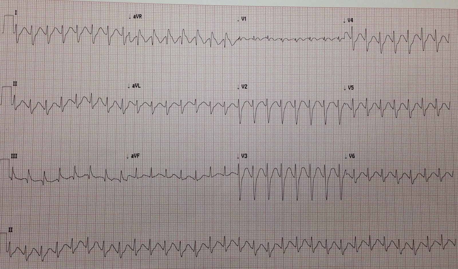
Wide complex tachycardia.  This is mostly monomorphic VT, but because V5 shows 2 narrow beats (which could be mistaken for fusion beats), this is polymorphic VT due to short episodes of bidirectional ventricular tachycardia.  Notice these beats do not occur while V2 and V3 are being recorded, unlike the next ECG.

Second recording, shortly after the first:

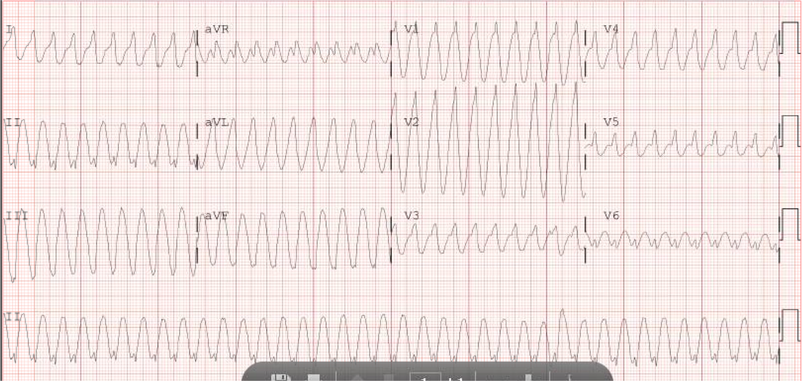
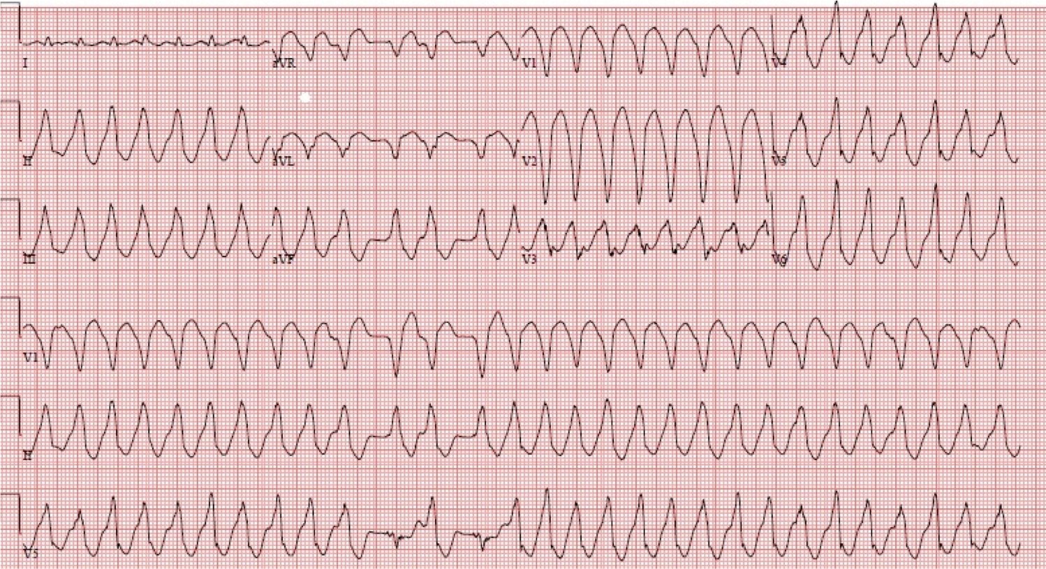
Here again we see Bidirectional Tachycardia, and it is more obvious because it occurs during the recording of V2 and V3, which show it more clearly than any lead except V5 (across the bottom),  which is by far the most diagnostic.

ED Course

He underwent synchronized cardioversion twice at 120 and 200 J with no response.  He maintained a wide complex tachycardia with a heart rate on the monitor up to 180, but with fewer perfused beats. He remained alert.

He was given metoprolol 15 mg IV with no response.  (Comment: I would consider this a small dose, and would give esmolol which is short acting and can be dialed up and stopped if there are adverse events).

In order to diminish the catecholamine surge thought to be driving his tachycardia, the patient was intubated and sedated with propofol.  This failed.

Overdrive pacing was attempted via his implanted pacemaker, and this also failed.

The rhythm was then managed with amiodarone (this failed).

He was given esmolol (this also failed).

Finally, he was given verapamil.

Following verapamil, the patient became very hypotensive until he became ashen and pulseless.  Chest compressions were started and he was defibrillated again at 200J, resulting in restoration of sinus rhythm at 30-40 bpm, with inadequate perfusion.

Atropine and pacing were instituted.  The patient stabilized, and he was able to be transferred out of the ED.

After a week in the hospital, he was discharged on amiodarone and nadolol after a benign stress test. Flecainide was discontinued.  There was consideration of future sympathetic denervation of the heart.

Catecholaminergic Polymorphic Ventricular Tachycardia

CPVT is one of the types of VT that occurs in a structually normal heart.  It is due to rare mutations (either inherited or spontaneous) of either the ryanodine receptor gene or the calsequestrin 2 gene.  Both in turn affect diastolic calcium release from the sarcoplasmic reticulum, and thus increase intracellular calcium levels (just as digoxin does), leading to delayed afterdepolarizations and triggered activity.  Just as with Digoxin, one of the primary dysrhythmias is Bidirectional Tachycardia.  

PMVT is characterized by episodic syncope during strenuous exercise or acute emotion in people without structural cardiac abnormalities.  It produces polymorphic VT, often with bidirectional VT, and can be well tolerated.

As mentioned above, some CPV are linked to mutations in the cardiac ryanodine receptor, also called the cardiac sarcoplasmic calcium release channel (which mediates the release of calcium from the sarcoplasmic reticulum, required for myocardial contraction).  Catecholamines can cause these ryanodine related channels to “leak” calcium in diastole and can trigger life threatening arrhythmias.

Further discussion of mechanism is beyond this blog (and beyond me!)

The mean age of onset is 7-12 years, so this is initially a pediatric problem.  However, onset may be as late as 40 years.  The first manifestation may be cardiac arrest.

The baseline ECG is normal.  There are multiple mechanisms involved, including re-entry and delayed afterdepolarizations due to calcium overload or changes in autonomic tone.  It generally cannot be induced in the EP lab.

VT is of polymorphic morphology or of bidirectional morphology, such as one sees in digoxin or aconite toxicity, with alternating QRS axis, or alternating LBBB/RBBB.

Further discussion is beyond the scope of this blog, except for emergent, and a bit on chronic, management.


ACUTE MANAGEMENT of Catecholaminergic Polymorphic VT

(there is little in the literature that I could find; the ACC/AHA guidelines on dysrhythmias have not, apparently, been updated since last published in 2006):

If it is a child with his/her first presentation, you may be presented only with polymorphic VT of unknown etiology.  If you can record an ECG in sinus, and there is a long QT, then you will know that it is not PMVT.  If you do not have that opportunity to record it, you will not know, but long QT is far more common than PMVT.  By the time of adulthood, the patient will probably be aware of the condition.  This is important, because the management of the two entities is very different.

If you are able to record an episode of sinus rhythm:

1. If there is a long QT, then the polymorphic VT is Torsade.

2.  If there is ischemia or evidence of cardiomyopathy on the ECG, then it is polymorphic VT due to ischemia or a structurally abnormal heart.

Here is a post on the management of Polymorphic VT NOT due to CPVT.

3. If the QRST is normal when in sinus, then strongly consider CPVT.

Or perhaps, as in this case, the patient is known to have CPVT.

Here are some management steps to take:

1. Cardioversion or defibrillation (depending on presence of pulses), but with beta blocker pre-treatment (if possible), as the shock itself can cause catecholamine surge, further aggravating the situation and causing early recurrence of VT.

2. Intravenous beta blockers should be administered.  Esmolol is a great choice, as it has a very short half-life and can be turned off if there are complications.  Dose: Esmolol can be given in a bolus of 500 mcg/kg (0.50 mg/kg), followed by a drip of 50 mcg/kg/min (0.05 mg/kg/min).  It can be rebolused repeatedly, as needed, each time increasing the infusion by 50 mcg/kg/min.

3. For recurrent VT in the absence of prolonged QT, amiodarone should be administered by bolus and drip.  Amiodarone has beta-blocking effects.

4. Verapamil is recommended based on studies in which it prevents exercise-induced PMVT (3, 4).  However, I haven’t found literature on its use in the acute situation.  Our electrophysiologist wrote that he would try it.  In this case, it appears to have made things worse, but that is the problem with isolated cases without control groups.


4.  Sedation, as in this case, to decrease sympathetic drive?  Or, class IIb in ACC/AHA guidelines for incessant VT is general anesthesia.

Avoid isoproterenol and overdrive pacing.  For polymorphic VT due to acquired long QT (Torsade de Pointe), isoproterenol is indicated to increase the sinus rate and prevent further torsade.  However, this makes CPVT worse.  Fortunately, Torsade is more common, but if you know the patient has CPVT, avoid isoproterenol.

Overdrive Pacing is listed as class IIb for incessant VT in the ACC/AHA guidelines, but this should not to be applied to CPVT. 

Overdrive pacing is used to prevent recurrence of pause-dependent PMVT (Torsade), which is worsened by bradycardia and improved by tachycardia).  The pathophysiology of CPVT is very different from Long QT syndrome.  CPVT, unlike Torsade, is worsened with tachycardia.   See this post for management of Torsade.

CHRONIC MANAGEMENT (these are a few therapies that the electrophysiologist may pursue)

1. Beta blockers

2. AICD consideration, especially if survivor of cardiac arrest.  ICD treatment without concomitant use of beta blockers
is dangerous because of the risk of electrical storm induced
by the adrenergic surge related to a shock.

3. Flecainide if refractory to above (it inhibits the cardiac ryanodine receptor)

4. Verapamil

5. Strictly avoid competitive sports

6. Sympathetic denervation if refractory to above

Missing just one dose of beta blocker can lead to a fatal or near fatal event

This is copied from the ACC/AHA guidelines (quote):

Supraventricular and ventricular arrhythmias are usually reproducibly induced by exercise stress when the heart rate reaches a threshold of 120 to 130 beats per minute. Isolated PVCs usually develop first and are followed shortly after by short runs of NSVT. If the patient continues to exercise, the duration of VT runs progressively increase and VT may become sustained. A beat-to-beat alternating QRS axis that rotates by 180°, “bidirectional VT,” is the typical pattern of CPVT-related arrhythmias. CPVT patients can also present with irregular polymorphic VT or VF.


Beta blockers prevent recurrences of syncope in the majority of patients, even if VT and SVT usually can still be elicited during exercise stress test. If syncope occurs in a patient taking a beta blocker, the implantation of an ICD is recommended. However, ICD therapy requires careful programming of the device to prevent needless therapy for nonsustained episodes of ventricular tachyarrhythmia.

References:

Zipes DP et al.  ACC/AHA Guidelines for management of patients with ventricular arrhythmias and sudden cardiac death.  JACC Vol. 48, No. 5, Sept. 2006:e247– e346

Pflaumer A.  Davis AM.  Guidelines for the diagnosis and management of
Catecholaminergic Polymorphic Ventricular
Tachycardia.  Heart, Lung, and Circulation 2012:21(2):96-100.

Rosso R, et al. Calcium channel blockers and beta-blockers versus beta-blockers alone for preventing exercise-induced arrhythmias in catecholaminergic polymorphic ventricular tachycardia. Heart Rhythm 2007;4:1149–1154.

Swan H, et al. Calcium channel antagonism reduces exercise-induced ventricular arrhythmias in catecholaminergic polymorphic ventricular tachycardia patients with RyR2 mutations. J Cardiovasc Electrophysiol 2005;16:162–166.  This article only comments on chronic management, not acute management.

  • Catecholaminergic Polymorphic VT
  • Polymorphic Ventricular Tachycardia
Steve Smith
onFebruary 6, 2015
  • Catecholaminergic Polymorphic VT
  • Polymorphic Ventricular Tachycardia
Share
Previous Article

Beware Automated Synchronization for Cardioversion!

Next Article

A Relatively Narrow Complex Tachycardia at a Rate of 180.

View Comments (9)
  1. Christopher
    on February 6, 2015

    Leenhardt 2012 (PMID: 23022705) reports that propafenone has also been shown to block RyR2 similar to flecainide (but only used in a single case report of resistant CPVT). They also reported that dantrolene is another possible option for resistant CPVT.

    They too lament the lack of therapeutic strategies, but pointed to ongoing research into RyR2 inhibitors.

    Reply
    1. Steve Smith
      on February 7, 2015

      Thanks, Christopher!

      Reply
  2. ECG Interpretation
    on February 6, 2015

    GREAT post on a less common but important entity. THANK YOU!

    Reply
    1. Steve Smith
      on February 7, 2015

      Thank you, Ken.

      Reply
  3. BendoMD
    on February 7, 2015

    This was very useful. I am a resident on cardiology.

    Reply
  4. Dr. Han Naung TUn
    on March 3, 2015

    Very nice presentation. Bravo.

    Reply
  5. Anonymous
    on January 9, 2017

    Thank you for this informational article! I actually have CPVT and have been trying to research and learn more about it myself.

    Reply
  6. Unknown
    on November 17, 2020

    Sorry I'm late to the game. Would this be an indication to try a stellate ganglion block? I've attached my notion of the idea, as taken from Josh Farkas in the IBCC blog for polymorphic VT storm (not saying that Farkas suggests it here. More asking, would this pathology or presentation be consistent with his pathway for VT storm (without QT prolongation aka not Torsades) https://emcrit.org/ibcc/storm/

    Reply
    1. Steve Smith
      on November 17, 2020

      I'm not sure, but I suspect not. I think that most of the catecholaminergic stimulation comes from the adrenal gland (epinephrine) rather than through direct ganglionic sympathetic stimulation.

      Reply

Leave a Comment Cancel

Your email address will not be published. Required fields are marked *

About

Steve Smith Avatar

Dr. Smith

faculty physician

Dr. Stephen W. Smith is a faculty physician in the Emergency Medicine Residency at Hennepin County Medical Center (HCMC) in Minneapolis, MN, and Professor of Emergency Medicine at the University of Minnesota.

Dr. Smith's Google Scholar Profile

Dr. Smith Articles on PubMed

Creative Commons License

This work is licensed under a Creative Commons Attribution-NonCommercial 4.0 International License.

Follow us on X (Twitter)

Follow @smithECGBlog
Follow @PendellM
Follow @ekgpress
Follow @AslangerE
Follow @ecgcases
Follow @PMcardioBot

Featured Posts

‘NSTEMI’ or reperfused OMI? And which lesion is the culprit?
Jesse McLaren
onFebruary 8, 2025
‘NSTEMI’ or reperfused OMI? And which lesion is the culprit?
Jesse McLaren
onFebruary 8, 2025
“A patient just arrived as a transfer for NSTEMI.”
Willy Frick
onDecember 7, 2023

Blog Archive

Labels

"inferior" ST depression (22) anterior STEMI equation (14) anterior T wave inversion (15) atrial flutter (28) AV block (17) AV dissociation (13) aVL (31) aVR (30) Benign T-wave Inversion (23) bradycardia (13) cardiac arrest (54) cardiogenic shock (15) computer (21) CT scan (13) de Winter's T-waves (25) Diffuse ST Depression (18) Dr. K. Wang (20) early repolarization (22) echocardiogram (24) false positive cath lab activation (45) False positive ECG (21) Formula for Early Repol vs. Anterior STEMI (39) hyperacute T-waves (80) Hyperacute T-waves LAD (24) hyperkalemia (63) hypokalemia (32) inferior hyperacute T-waves (24) Inferior MI subtle (31) inferoposterior STEMI (15) LAD occlusion (107) LAD occlusion vs. benign early repolarization (43) LAD OMI missed (18) LAD reperfusion (15) LBBB (54) lead misplacement (20) left bundle branch block (16) left main (22) long QT (45) LV aneurysm (35) LVH (41) LVH and OMI (13) missed OMI (30) missed STEMI (23) modified sgarbossa criteria (37) Morphine (13) Myocarditis (20) Normal ECG by computer algorithm (56) OMI (147) paced rhythm (24) Pediatric (15) pericarditis (25) PM Cardio Queen of Hearts (66) posterior leads (22) posterior OMI (44) posterior reperfusion T-waves (16) posterior STEMI (26) Precordial swirl (32) prehospital ECG (19) pseudoinfarction (23) pseudonormalization (25) PseudoSTEMI (36) pulmonary embolism (28) PVC (15) QOH (14) RBBB (49) RBBB with LAFB (Left Anterior Fascicular Block) (20) reperfusion (21) RVH (13) RV MI (20) saddleback STE (13) serial ECG (34) spontaneous reperfusion (17) ST depression (40) subacute MI (13) subendocardial ischemia (16) subtle (41) Subtle LAD (51) Subtle LAD Occlusion (55) Subtle STE (14) Syncope (33) T-Wave inversion (26) takotsubo (24) terminal QRS distortion (16) Third (3rd) degree AV block (13) torsade (14) transient ST elevation (19) U-waves (23) ultrasound (19) unstable angina (27) Ventricular Fibrillation (14) ventricular tachycardia (29) Wellens' syndrome (31) wide complex (16) Wide complex tachycardia (43) wide QRS (13) wide_complex_tachycardia (25) wolff parkinson white WPW (24) wraparound LAD (15) young (18) Young Women (17)

Read Next

  • sinus tach misinterpreted as SVT
  • Sinus Tachycardia Extreme
  • Supraventricular Tachycardia (PSVT)
5 Min Read
Steve Smith
onFebruary 8, 2015

A Relatively Narrow Complex Tachycardia at a Rate of 180.

I received a text message with this image: “Cardioversion didn’t work.  Any thoughts?” What do you think?  The…
  • Excessively discordant ST elevation
  • LBBB
  • STEMI-equivalent
1 Min Read
Steve Smith
onFebruary 11, 2015

New Left Bundle Branch Block (LBBB) and Dyspnea

It is important to remember that the latest (2013) ACC/AHA STEMI guidelines removed New Left Bundle Branch Block (LBBB) as an indication for…
  • Adenosine
  • AVRT: AV Reciprocating Tachycardia (through accessory pathway)
  • Dual AV nodal pathways
  • wide_complex_tachycardia
6 Min Read
Steve Smith
onFebruary 16, 2015

Wide Complex Tachycardia in a 20 something.

This was sent by a former resident.  He will remain anonymous because his identity could compromise patient confidentiality. Case A…
Dr. Smith’s ECG Blog Dr. Smith’s ECG Blog
Expert ECG Interpretation and Emergency Cardiology Education
© 2025 — Dr. Smith's ECG Blog. All Rights Reserved.