Dr. Smith's ECG Blog

Instructive ECGs in Emergency Medicine Clinical Content

Associate Editors:
— Pendell Meyers & Ken Grauer (2018)
— Jesse McLaren & Emre Aslanger (2022)
— Willy Frick (2024)

editors

Pseudo-Wellens’ Syndrome due to Left Ventricular Hypertrophy (LVH)


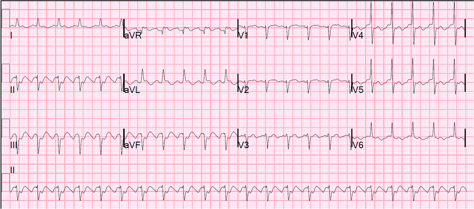
A 52 year old male presented with epigastric pain, continuing in the emergency department.  He has a history of hypertension, and is on amlodipine.  Thinking that epigastric pain might be an anginal equivalent, this ECG was recorded:

There is LVH.  There are T-wave inversions in aVL and V3-V6. 


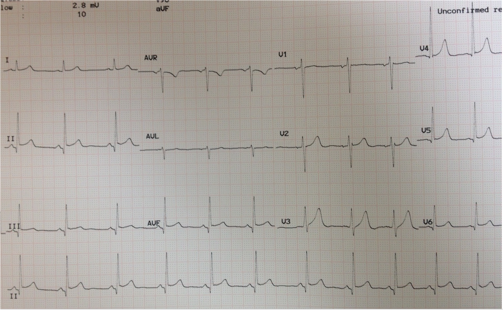
The physicians were worried about Wellens’ syndrome and treated for ACS.  He was placed on a nitroglycerine drip.  Initial troponin was negative.  A followup ECG was done 3 hours later.

Perhaps slightly less T-wave inversion.  Nonspecific.


He was admitted.  All troponins were normal.  A formal echo showed concentric LVH and there was also a subtle abnormality which the echocardiographer thought suspicious for LAD distribution ischemia.  So he underwent a coronary angiogram which was completely normal.

This is not Wellens’.  Why?

1.  LVH frequently causes T-wave inversion which mimics Wellens’

2.  In Wellens’, the chest pain is nearly always resolved by the time of the ECG.  It is reperfusion that results in the T-wave inversion and the pain is thus resolved by this time.

3.  Wellens’ is defined by chest pain and ECG abnormalities.  Though Acute MI can present as epigastric pain, it is not at all common and T-wave inversion in the setting of epigastric pain is not high risk for ACS.

4.  T-wave inversion in Wellens’ is primarily V2-V4.  When T-wave inversion is V3-V6, one should think more about LVH or Benign T-wave Inversion (this post on Benign T-wave Inversion is worth reading)

Previous Article

Dynamic inferior MI: STEMI or Non-STEMI? Does it matter?

Next Article

What is the Diagnosis?

View Comments (12)
    1. For me, I simply recognize the pattern. I have seen so many.

      For others, I just recommend that you think about LVH and, if there is LVH, you must suspect a false positive, as in this case.

  1. Hello.. If LVH is diagnosed by lead aVL>11mm criteria n chest leads amplitude being normal, would those wellens waves still be called wallens or pseudo wellens? Thx

    1. No simple answer. Criteria are not very good for LVH. And much LVH does not have repolarization abnormalities, so this does not apply to all LVH. To know this is not Wellens', you need the totality of the presentation: Not chest pain (epigastric), still ongoing, T-wave inversion V3-V6, AND LVH. No one thing will give you the answer.

    1. I didn't use criteria. Just looked at it and knew. But if you do apply the best criteria (recently published), it is LVH.

      New LVH criteria: sum of the amplitude of the deepest S wave in any lead plus the S wave in lead V4. If the deepest S wave is in V4, then double that value. If the total is greater than 2.3 in women and
      greater than 2.8 in men then left ventricular hypertrophy is diagnosed.

      Peguero JG et al. Electrocardiographic criteria for the diagnosis of left ventricular Hypertrophy. JACC 69(23):1694-1703; April 4, 2017

  2. Maximilian.karla@gmail.com

    Dear Dr. Smith,
    I would like to ask you if you would consider Wellen's syndrome or Pseudo Wellen's syndrome with this ecg:

    https://photos.app.goo.gl/DzGxKRwF2myNo4DS6

    Background information: 70 years old, male, no chest pain but chest pressure ongoing for ~40h, 2 former mci, troponin negative?

    Thank you!

    1. ECG Interpretation

      @ Maximillian — I have reviewed your tracing at the above link. I would not call this Wellens or pseudo-Wellens — because I think the ECG appearance is quite different from what I’d expected with true Wellens Syndrome. FIRST — the history (sounds like there is ONGOING chest pressure for ~40 minutes) is not typical (ie, with Wellens Syndrome, as per Dr. Smith — there is typically a history of prior chest discomfort, but none by the time the ECG is obtained). SECOND — we NEED to see the prior ECG, given that this patient has had 2 prior MIs. True Wellens Syndrome entails acute findings — and this wouldn’t be the case if much of what we see on your tracing are longstanding findings. Otherwise — there is MARKED LVH (overlap of a very tall R wave in lead V5 — that to me looks to be at least 23-24mm tall + a R in V6 = 16mm, with ST-T wave changes in these 2 leads perfectly consistent with LV “strain”. There is also a peculiar notching of the T wave in leads V2-V4 (hard to rule out possibility of U waves here …) — which together with clear ST segment flattening, with slight ST depression + biphasic T waves in a number of leads could be: i) electrolyte abnormality (ie, low K+ and/or low Mg++); ii) drug effect; iii) longstanding ischemia; iv) LV “strain” (at least in certain leads). The findings certainly aren’t localized, as I’d expect them to be with Wellens or pseudoWellens. Hope that answers your question. THANKS for your comment! — 🙂

  3. Kaushik Virivinti

    Dear dr smith I would like you to confirm if the patient's ecg below has wellens waves
    History 50 year old female, recent on set of hypertension,diabetic, complained about moderately severe arm pain ecg was taken 4-5 days after the incident
    My friend says is it wellens pattern , I dont agree with him as it should have progressed to complete inversion of t waves by the time ecg was taken please help us out

Leave a Comment

Your email address will not be published. Required fields are marked *