In the last post, I showed:
Ten (10) Examples of Hyperacute T-waves in Lead V2 (a few in V3), due to acute LAD occlusion
Today, 10 examples of inferior hyperacute T-waves.
Details in every case make it even better, so click on the link!!
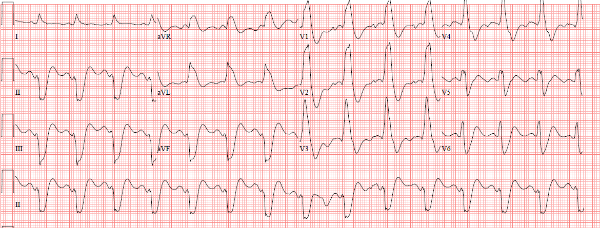
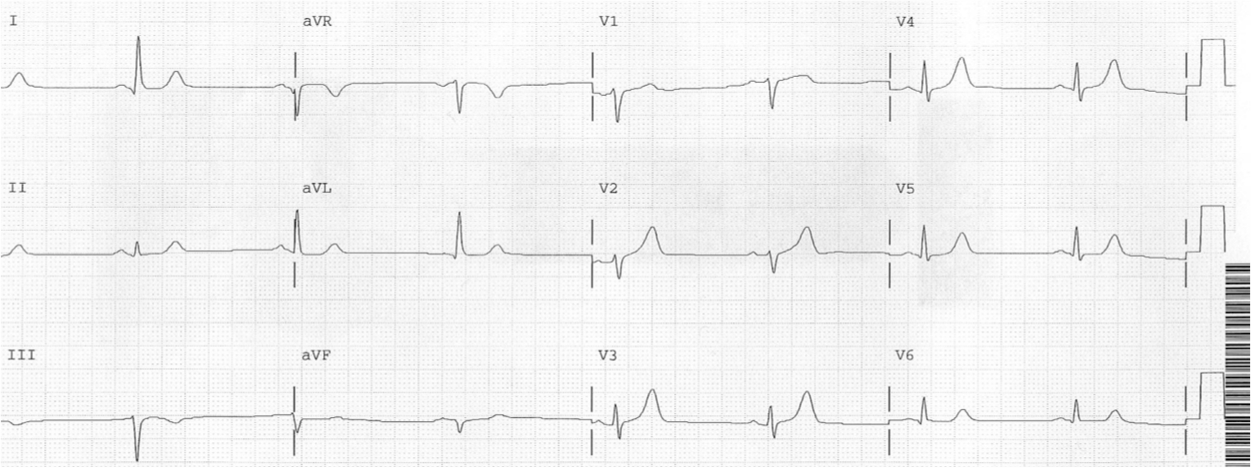
Case 1.
Chest Pain in a 61 year old male. Inferior STEMI without inferior ST elevation.
 |
| In this case, the followup ECG is diagnostic because of lead V1 |
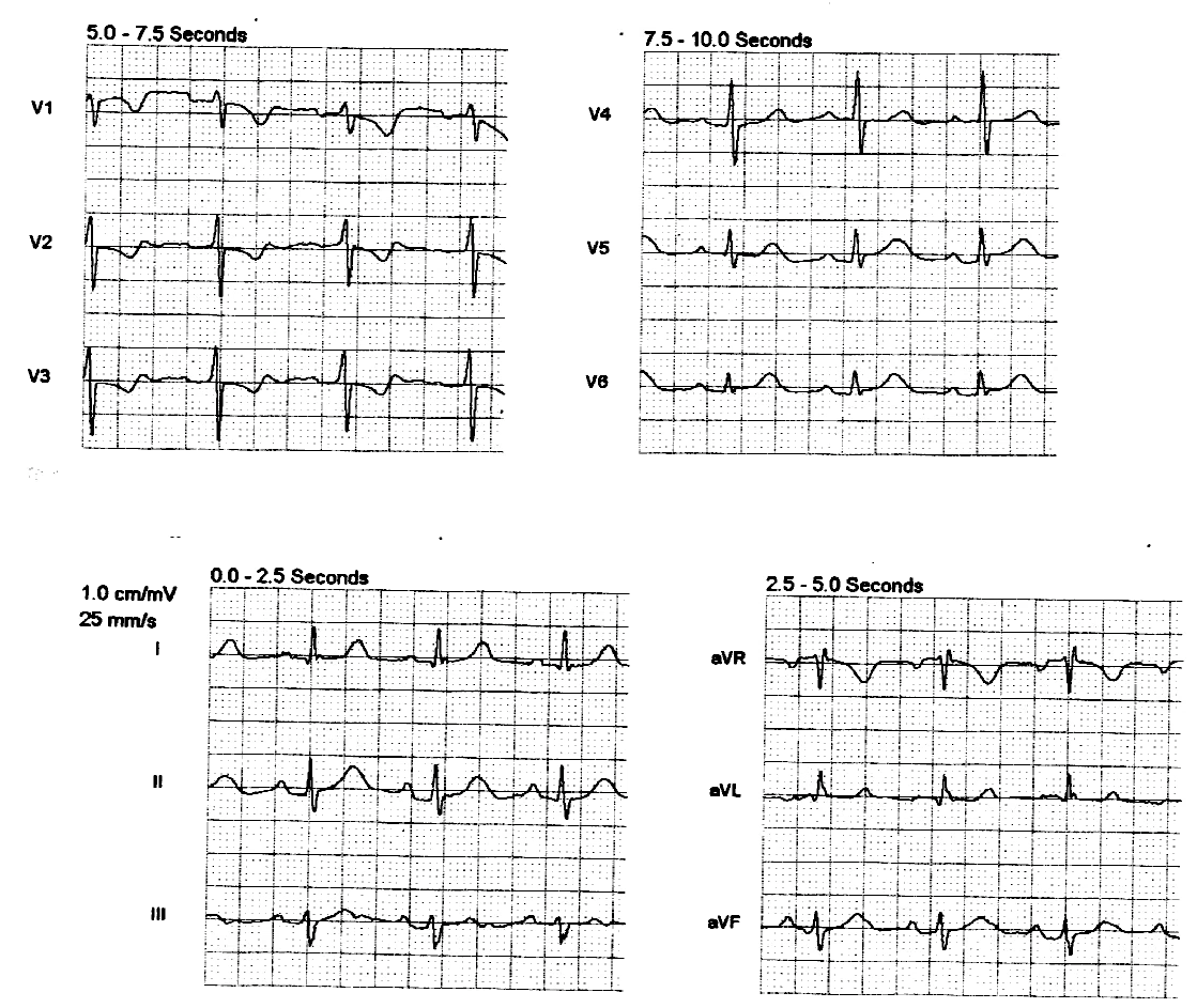
Case 2.
Inferior Hyperacute T-waves

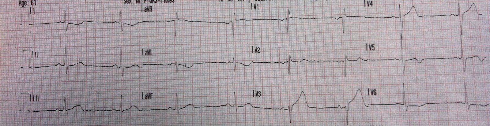
Case 3.
Chest Pain in a Male in his 20’s; Inferior ST elevation: Inferior lead “early repol” diagnosed. Is it?
 |
| This case was missed. Read about it. |
Case 4.
Series of Prehospital ECGs Showing Reperfusion
 |
| This has an incredible sequence of ECGs! |
Case 5.
The development of an inferior-posterior STEMI, from prehospital to hospital

Case 6.
Friday’s post produced skeptics…..
 |
| This case made a lot of readers angry, denying that these are hyperacute Ts |
Case 7.
A Case of Clinical Unstable Angina in the ED
 |
| This case shows how hyperacute is only relative to the baseline T-waves. Also, the critical role of reciprocal ST depression and T-wave inversion in aVL. |
Case 8.
Is this STEMI? Pattern Recognition is Key
 |
| Go to the link, and this case shows an inferior pseudoSTEMI for contrast |
Case 9.
Inferior hyperacute T-waves. The clue is T-wave inversion in aVL. Serial ECGs evolve to ST Elevation.

Case 10.
Subtle Inferoposterolateral STEMI