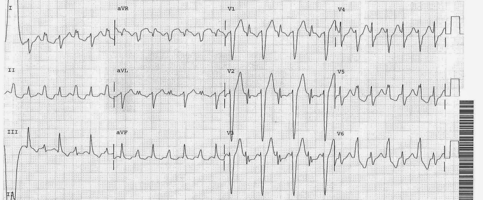
A 50 yo male presented with chest pain. This ECG was recorded at 0415.
 |
|
|
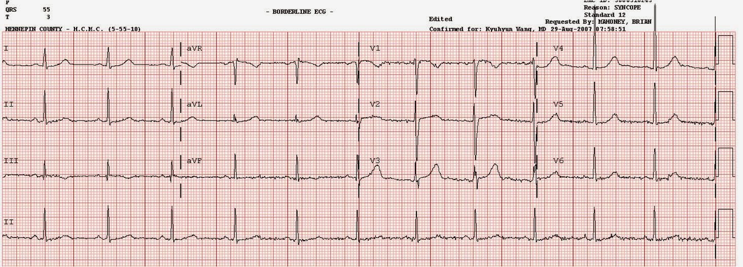
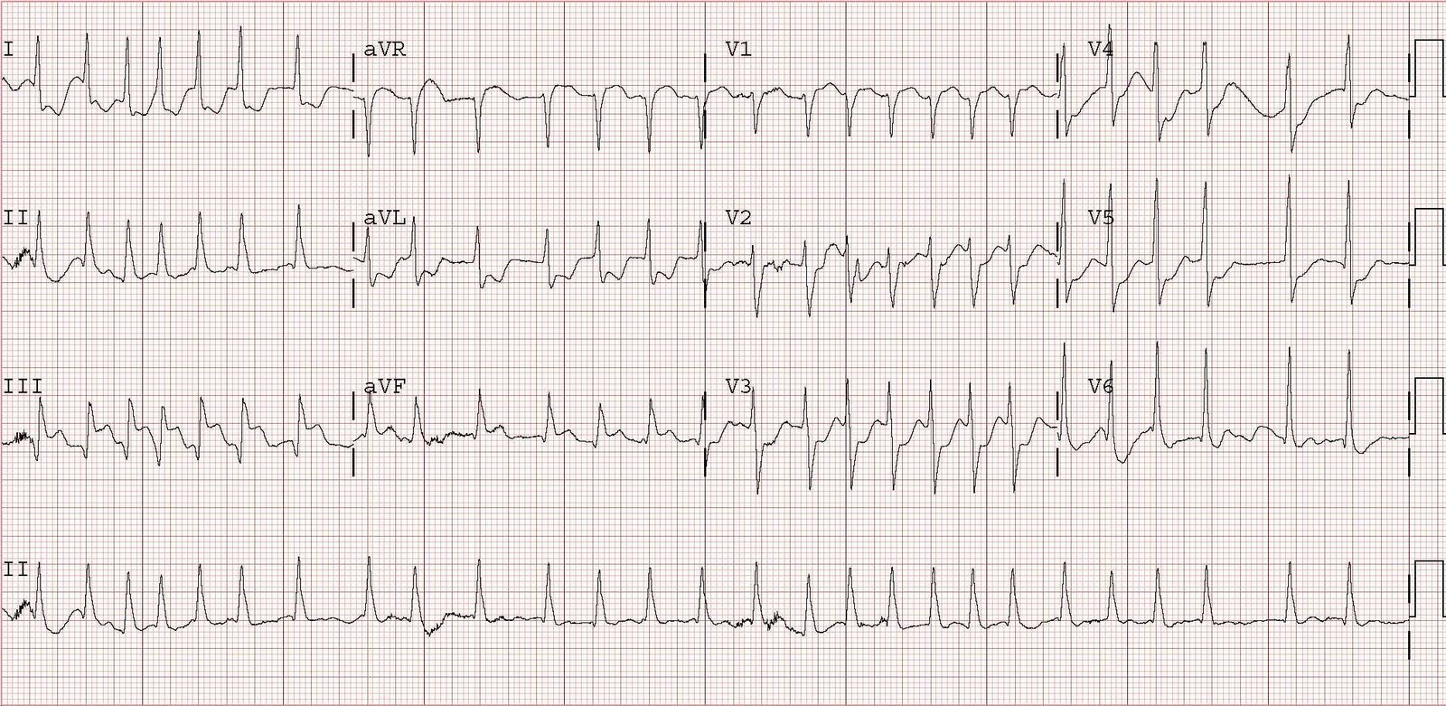
The occlusion was not appreciated by the treating physicians, and another ECG was recorded at 0457:
 |
|
|
The notching in V3 is also known as “Cabrera’s sign” (prominent notching of at least 40 msec in the ascending limb of the S-wave in any of leads V3-V5).
The patient was taken to the cath lab and a 100% LAD occlusion was opened.
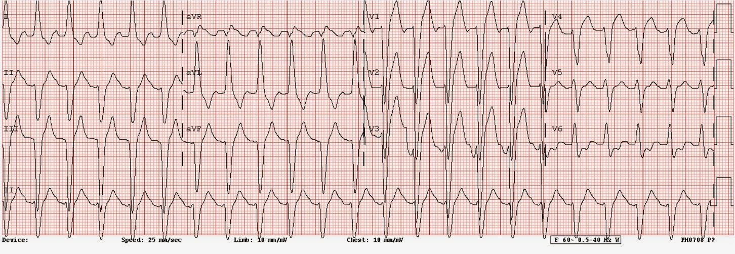
Here is the ECG after reperfusion:
 |
|
|
Such T-wave inversion is a frequent sign of reperfusion even in LBBB, and when seen alone (without the preceding ECGs diagnostic of STEMI) is a common sign of NSTEMI. T-wave concordance can be normal, so it is not a very specific nor sensitive sign of ischemia. But it should raise your suspicion. In the context of this case and the preceding ECGs, it is diagnostic of reperfusion and is definitely the result of ACS.