This is an old post from June 2009 that has not received much attention, but should. Take a look!
This 80 yo woman had been increasingly lethargic for 2 days, and presented hypotensive (SBP =70), pale, and tachycardic. She had not been complaining of chest pain. It was uncertain whether she had chronic atrial fibrillation or not. She was afebrile. Here is the initial ECG.
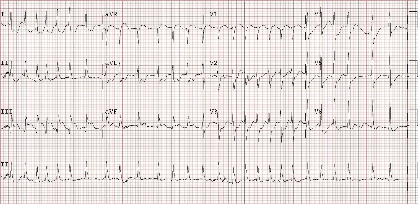
There is an irregularly irregularly rhythm (atrial fibrillation) with a very fast ventricular response. There is an injury pattern, with ST elevation in II, III, aVF, reciprocal ST depression in I and aVL, and ST depression of posterior injury in precordial leads.
We did not activate the cath lab. We suspected GI bleed and this was confirmed with blood on rectal exam. An ultrasound of the heart showed hyperdynamic function and the inferior vena cava (IVC) confirmed low central venous pressure (IVC was flat). Had this been a primary cardiac event, the CVP would be high and the IVC distended, and the patient might have also been in pulmonary edema.
She was given blood and fluids until the bedside ultrasound showed good central venous pressure (distended inferior vena cava), but she remained hypotensive and tachycardic, and the ST elevation did not resolve. Thus, we sedated her and electrically cardioverted at 200J (biphasic), but this was unsuccessful x 3. We then intubated her and started an infusion of amiodarone 300 mg IV, but with no improvement, and a subsequent cardioversion was again unsuccessful. We then loaded her with 500 mcg/kg of esmolol and started her on a 50 mcg/kg/min drip, after which a fifth cardioversion was successful, and resulted in the second ECG shown here:

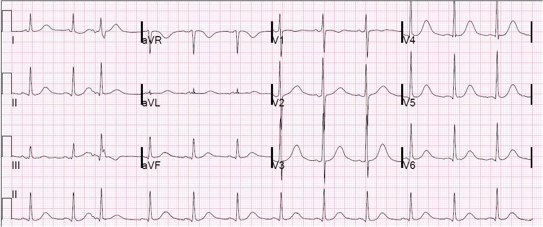
| The rhythm is sinus (with a couple PACs), rate normal, and all ST elevation and depression is now resolved. |
The blood pressure came up immediately. Hemoglobin returned at 7 and WBC count at 29,000. The troponin peaked at 19, and there was a subsequent inferior wall motion abnormality. EGD showed duodenal ulcers and erosions which had stopped bleeding.
A stress sestamibi showed no inducible ischemia, so no cath was done. Whether there was thrombus in the infarct-related artery, or whether this was only demand ischemia (Type II MI) is uncertain.
Convert atrial fibrillation with a rapid response when the patient is unstable; any injury pattern on the ECG constitutes instability.
Though demand ischemia usually shows as ST depression (or nonspecific findings) on the ECG, it may occasionally present with injury (ST elevation).