A young man was in a rollover Motor Vehicle Collision, with a chest laceration and chest pain.
He underwent a CT scan of the chest:
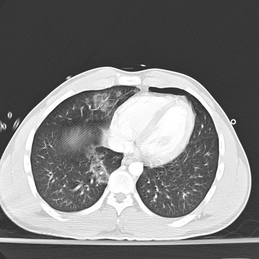 |
| There is a very small pneumothorax anterior to the heart |
This image is from slightly lower down:
 |
| There is a bit of air in the pericardium |
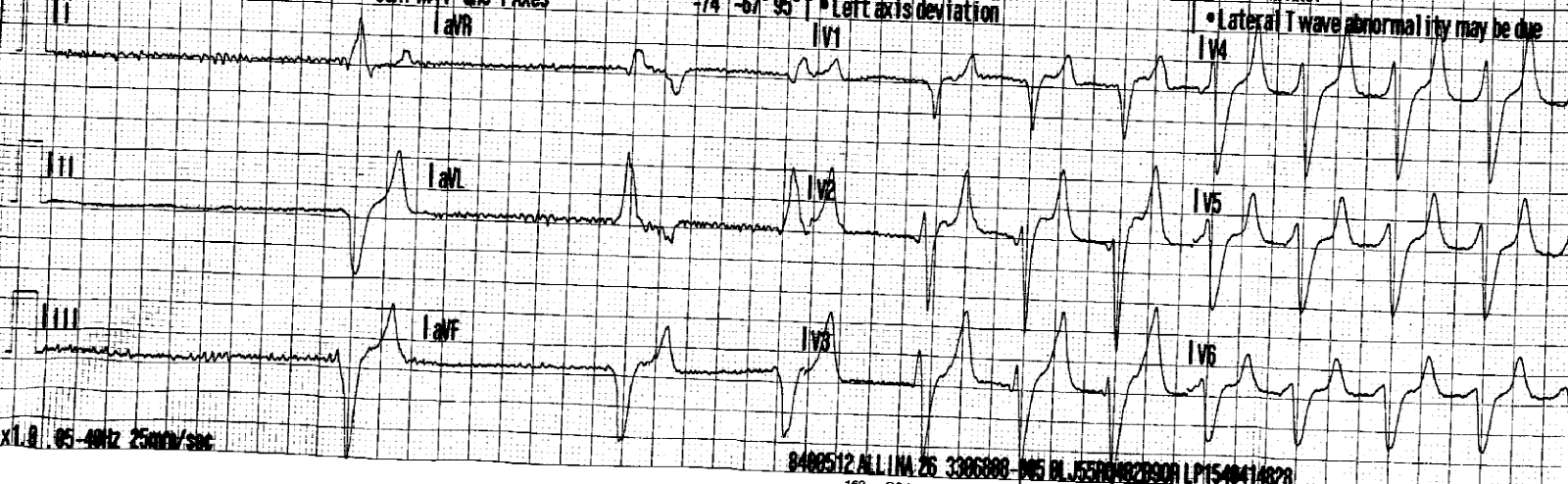
A troponin returned elevated, so this ECG was recorded:
 |
| There is some minimal ST elevation in inferior and lateral leads. This is consistent with percarditis. The troponin shows that there is some myocardial damage. Is it myocardial contusion, or myo-pericarditis? Or pericarditis as a result of myocardial contusion? |
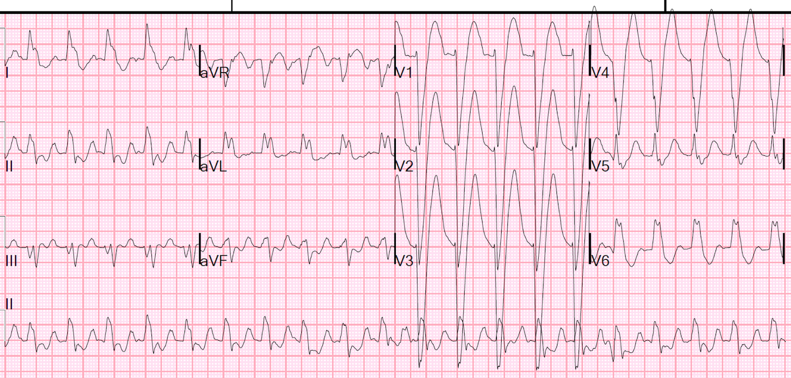
6 hours later:
 |
| There is diffuse ST elevation without large T-waves (low T-wave to ST segment ratio, which is typical of pericarditis vs. STEMI). There is PR depression. There is also “Spodick’s sign,” (downsloping TP segment); whether this is or is not a highly accurate sign of pericarditis is uncertain. |
The next morning
 |
| There is increased ST elevation |
Troponin Profile: