A middle-aged woman presented with what is described as a burning feeling in her chest which the physician said was “very atypical.” It did occur during exercise and radiated to both wrists.
Here is the first ECG:
 |
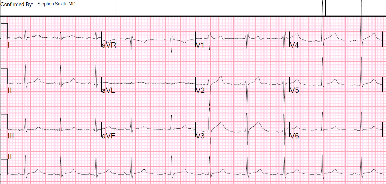
| Sinus rhythm.
Computerized QTc is 437. There is some ST Elevation in II and aVF but without reciprocal ST depression in aVL. Precordial leads also have ST Elevation: Is this normal variant or is it Ischemic ST Elevation? There is upward concavity in all leads, suggesting normality. But upward concavity is seen in all of leads V2-V6 in almost 50% of LAD occlusion. There is no ST depression, Q-waves, or T-wave inversion. |
The first impression of the clinicians was “pericarditis” because of the diffuse ST elevation.
The computer algorithm might say: “Diffuse ST elevation, consider pericarditis, early repolarization, or myocardial infarction.” I don’t know what it actually said.
Early repol vs. LAD occlusion
Should we use the LAD-Early Repol calculator?
If you did, and it was negative, it would likely be a false negative. Why?
There is Terminal QRS distortion in V3, which is not a finding of normal variant ST elevation.
What is Terminal QRS distortion?
Terminal QRS distortion is the absence of both an S-wave or a J-wave in either V2 or V3. It is not seen in early repolarization, or is very rare. In the right clinical context, and in the presence of non-diagnostic ST elevation, it is highly suspicious for coronary occlusion.
They did apply the formula, using these measurements: 1.5 mm for STE at 60 ms after the J-point in lead V3, QTc of 437, and R-wave amplitude in V4 of 13. (I would have used 2, 437, 14)
Their numbers resulted in 23.34 (very close to 23.4, but technically negative. I recommend that anything above 22.0 be investigated further)
My measurements would have resulted in 23.6, also very close but positive.
Pericarditis vs. LAD occlusion
I always say “You diagnose pericarditis at your (and your patient’s) peril.”
Why is this not pericarditis:
1. ST vector: The ST vector in pericarditis should be lateral and inferior and only slightly anterior. The vector here is towards V3.
2. Large T-waves: in pericarditis, the ST elevation is much more pronounced than the T-wave. Here the T-wave is more pronounced, hyperactute.
3. No diagnostic PR depression.
4. Notice there is a Spodick’s sign in V3-V5. But this is a worthless sign (see this recent post).
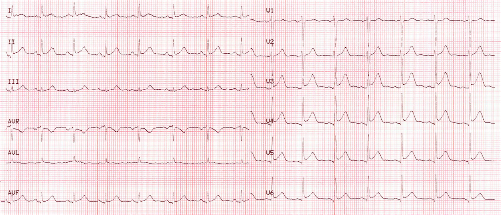
They recorded 2 more ECGs at unknown intervals:
2nd:
 |
| Perhaps some increase in STE |
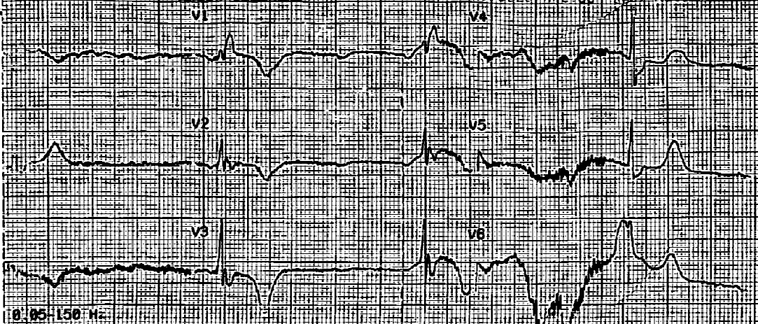
3rd:
 |
| There is slightly increasing ST Elevation |
Fortunately, the troponin came back slightly elevated, and fortunately they did not not attribute that elevation to myocarditis.
The patient was taken to angiography and found to have a 99% thrombotic LAD occlusion.