This post is written by Jack Cahill. He is an outstanding medical student and ECG reader. Some edits by Willy Frick and Smith.
A woman in her 50s, with hypertension, osteoarthritis, asthma, vitamin D deficiency and two previous transient ischaemic attacks, presented to the Emergency Department with 24 hours of chest pain. The documented history stated she had experienced chest symptoms for 1 week. Her observations were normal except for marked hypertension (BP ~ 170/100).
An ECG was obtained shortly after arrival:
ECG 1
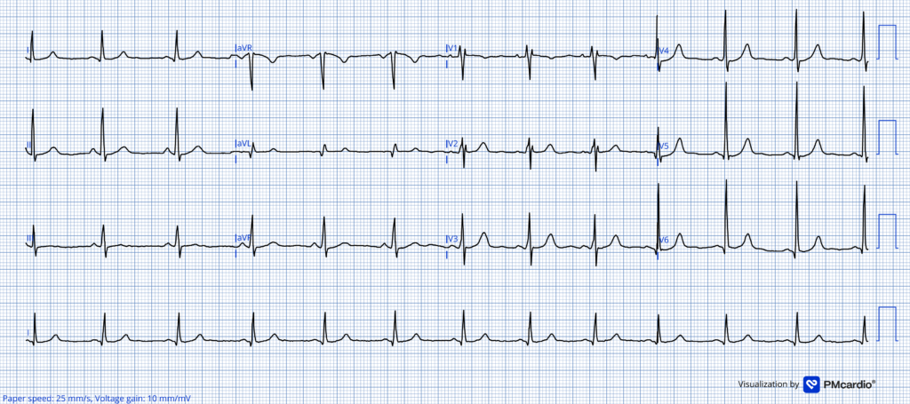
What do you think?
= = =
The ECG appears unremarkable. There is no obvious evidence of ischaemia, to me. Two high-sensitivity troponin assays were taken — only 50 minutes apart, both were “negative”. With apparently reassuring biomarkers and an ECG interpreted as normal, no further tests of ECGs were taken, and she was discharged home with simple analgesia and treatment for presumed reflux.
Smith: (details were unfortunately not in the chart). The only truly “negative” troponin is one that is undetectable. One should always report the exact values and the units. Without that data, we don’t know if this was a truly negative troponin.
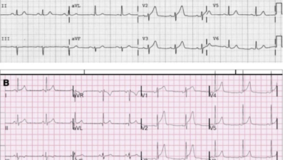
Unfortunately, her symptoms did not resolve. They worsened. She returned to the ED 5 hours later with ongoing “unbearable” chest pain. Her repeat ECG is shown below:
ECG 2
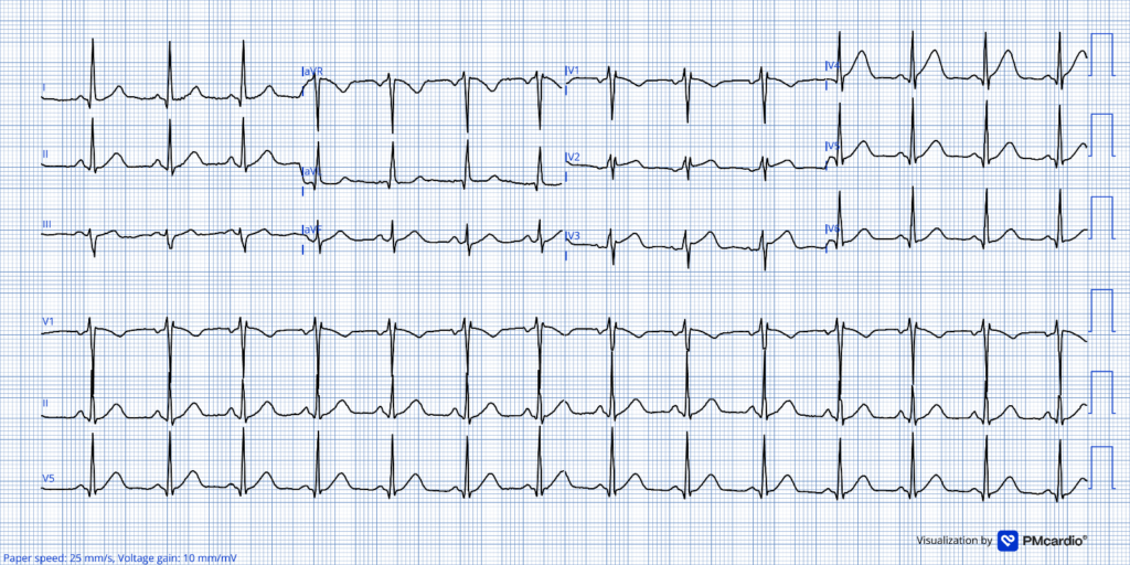
What do you think now?
= = =
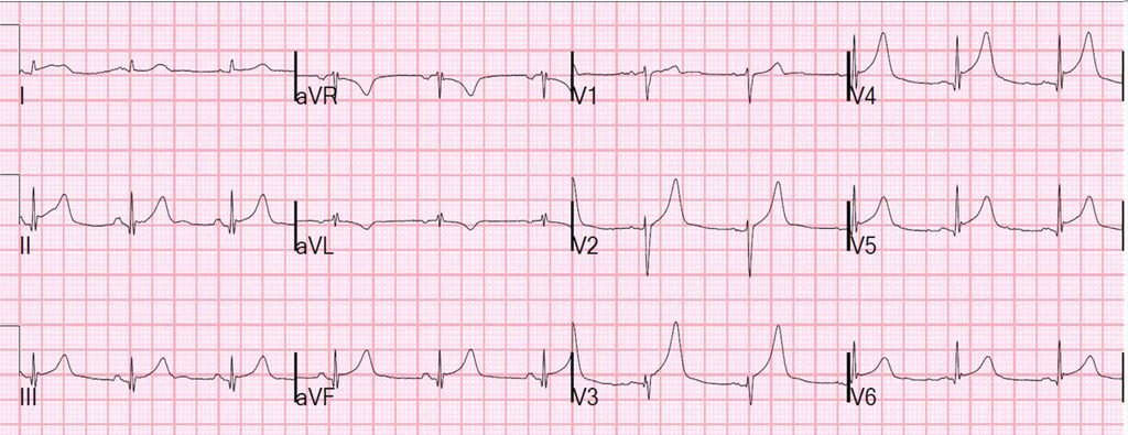
There is now a substantial increase in T-wave amplitude and area, disproportionate to the preceding QRS complexes. These are hyperacute T waves, most prominent in V4, but arguably present across both the inferior and anterolateral leads, particularly when compared with the previous ECG.
Smith: there is also a new Q-wave in lead V4 and slightly wider and deeper Q-waves in V5-6.
This is diagnostic of an acute LAD occlusion, most likely involving a wraparound LAD supplying the inferior apex.
This was not interpreted as STEMI/OMI and no immediate action was taken.
Smith: Here is the PMcardio Queen of Hearts AI ECG Model:
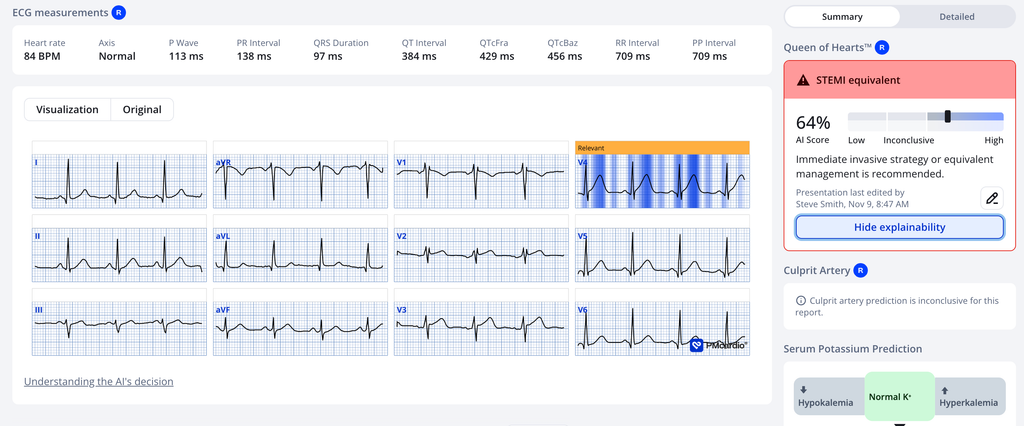
Smith: Our new model, which is not yet available, states that the differential diagnosis also includes “early repolarization”.
In spite of what they thought was a negative ECG, her initial troponin I returned at 250 ng/L.
The repeat, not measured until 12 hours later (!) rose dramatically to 48,000 ng/L, confirming extensive myocardial injury. She was treated for ACS and transferred to a PCI center.
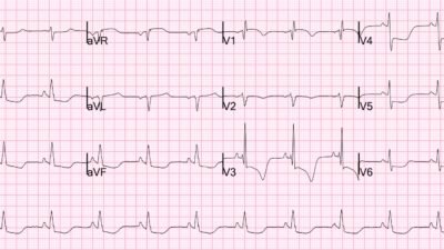
Repeat ECG obtained on arrival demonstrated evolving reperfusion changes, with deflation of the hyperacute T waves and the development of terminal T wave inversion across the anterolateral and inferior leads:
ECG 3
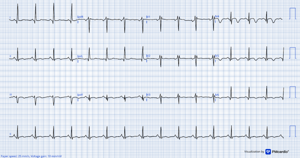
Coronary angiography performed the following day:
- Left main: Unobstructed.
- Left anterior descending: Type IV LAD, TIMI 3 flow, with an abrupt step-change in vessel calibre within the mid-vessel, suggestive of SCAD (spontaneous coronary artery dissection).
- Left circumflex: Dominant and unobstructed.
- Right coronary artery: Non-dominant, small calibre vessel.
No intracoronary imaging was performed and PCI was not undertaken. Echocardiography revealed left ventricular systolic dysfunction with apical ballooning and an akinetic apex.
Although this appearance resembles Takotsubo syndrome (stress cardiomyopathy), the coronary findings provide a different explanation. Angiography must always be performed before diagnosing takotsubo. A type IV LAD (wraparound LAD) supplies both the apex and the inferior wall; transient occlusion due to SCAD can therefore produce an extensive apical infarction with a classic ballooning appearance.
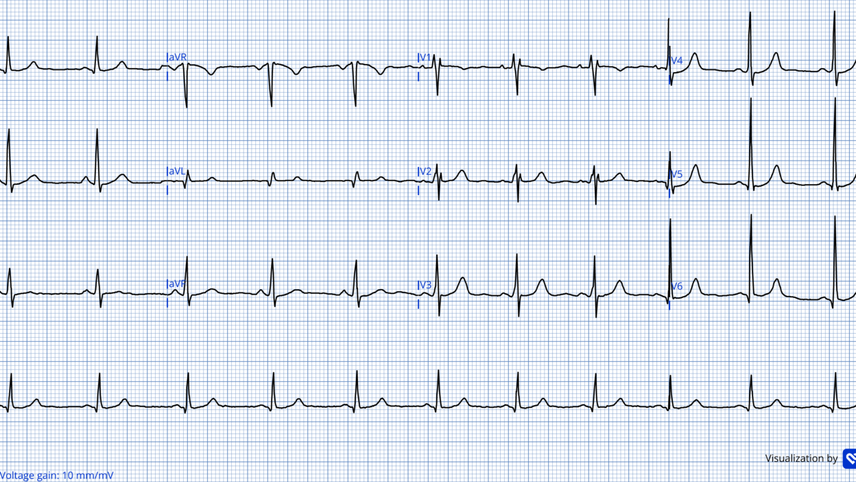
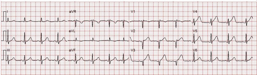
This was the ECG after coronary angiography:
ECG 4
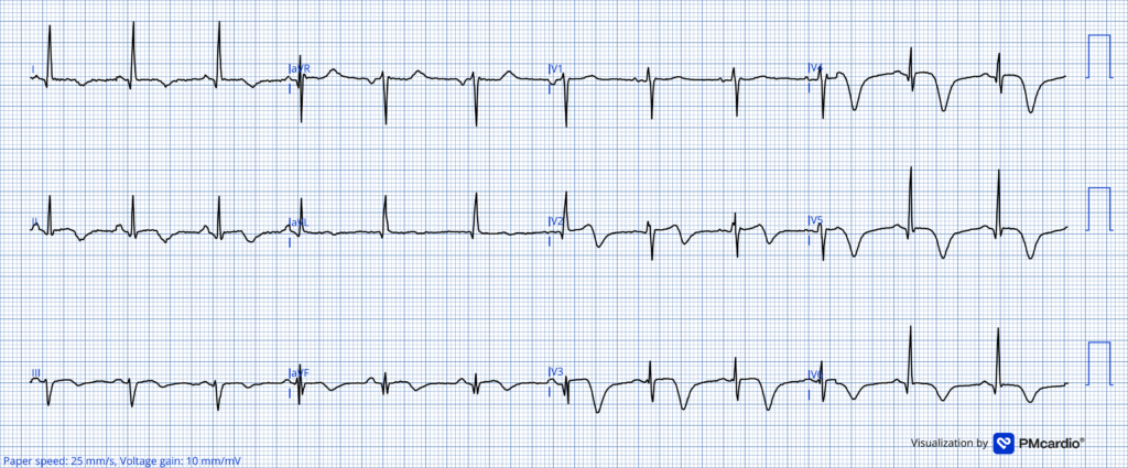
Ongoing anterolateral-apical reperfusion.
= = =
What does the Queen say about this one?
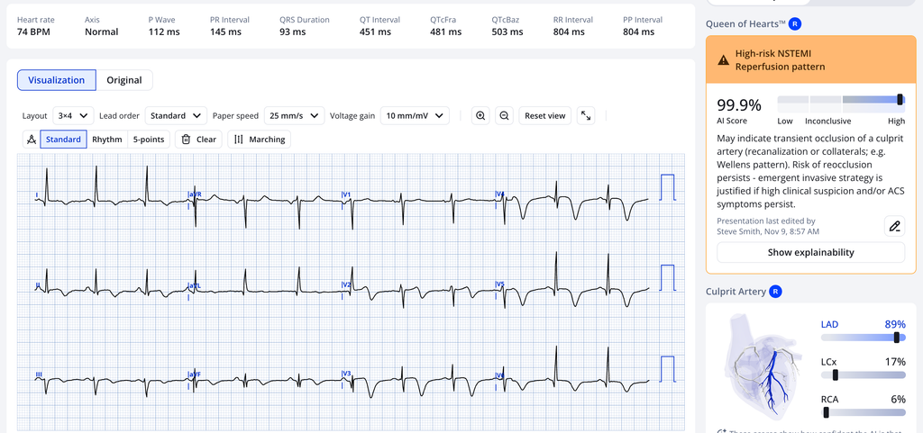
She agrees with reperfusion, with 99.9% certainty!
The overall diagnosis was: ‘Spontaneous Coronary Artery Dissection’.
= = =
Discussion
Women remain at higher risk of delayed or missed ACS diagnosis and contribute to experience worse outcomes following MI. In this case, the history documented was factually incorrect, which lowered suspicion for acute MI, potentially delaying care further than it already was. Prolonged or intermittent symptoms should not reassure you. Unstable coronary pathology may evolve through repeated episodes of occlusion and spontaneous reperfusion…
…Acute coronary occlusion is a dynamic process. The symptoms, ECG, troponin and stability of a patient will change as an infarct progresses. In these cases, time is of the essence — don’t wait for troponin or STEMI criteria before activating the Cath lab!
- SCAD is an under-recognized cause of ACS. SCAD results from disruption within the coronary vessel wall. SCAD is proposed to result primarily from expansion of an intramural haematoma that compresses the true coronary lumen.
- The intramural haematoma may arise following an intimal tear or from spontaneous bleeding within the vessel wall. Superimposed thrombus from the intimal injury may also compromise distal flow.
- In SCAD, coronary flow may fluctuate as a haematoma propagates or decompresses. Thus, by the time of coronary angiography, the patient may have an OMI(+) and/or STEMI (+) ECG, but TIMI 3 flow – which we see in ~ 20% of STEMI cases.
- Smith: The troponin of 48,000 is far too high for takotsubo. See data below. In an article (J Electrocardiol this year, see reference below), peak troponin I levels in takotsubo presenting without ST Elevation (which reflects this ECG) were median 0.59 ng/mL (IQR: 0.22-1.71) and initial were 0.27 (IQR: 0.09-0.99).
Perhaps the most important investigation omitted during the initial assessment, was not another blood test, but another ECG? Serial ECGs frequently demonstrate evolving ischaemia, long before biomarkers become unequivocally abnormal.
The echocardiographic appearance in this case also illustrates a diagnostic pitfall; A wraparound LAD can mimic takotsubo cardiomyopathy. Angiography remains essential before attributing this to stress cardiomyopathy.
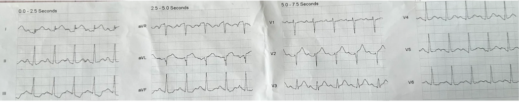
Smith: But ECGs also evolve in takotsubo, and it is often after T-wave inversion that they become much more recognizable because of the bizarrely long QT. Example: This ECG is nearly pathognomonic. What is it? (hint: it’s not Wellens’)
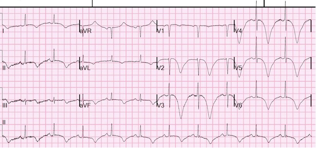
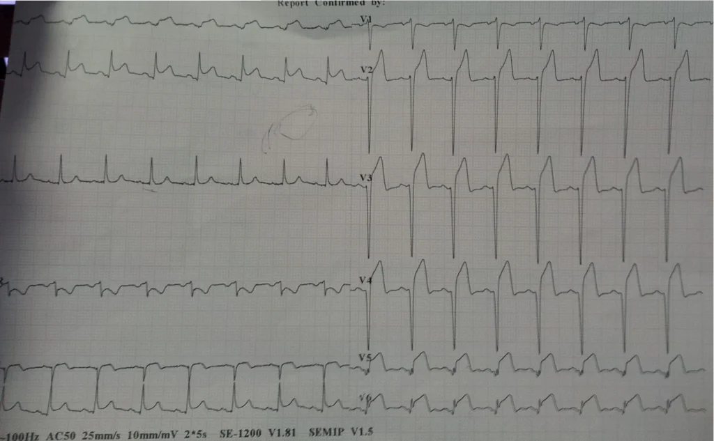
More cases here illustrating the difficulty of attributing apical ballooning to takotsubo, some of them below:
https://drsmithsecgblog.com/?s=wraparound+takotsubo
= = =
= = =
3. A 40-something woman with acute pulmonary edema — see the Speckle Tracking echocardiogram.
= = =
Some info on troponins on this post:
Troponins
The high sensitivity troponin I peaked at 9,324 ng/L
(approximately equivalent to 9.32 ng/mL by 4th generation and older assays.)
This is important because troponin I elevations in takotsubo are not that high. In a recent article (J Electrocardiol this year, see reference below), peak troponin I levels in takotsubo presenting without ST Elevation (which reflects this ECG) were median 0.59 ng/mL (IQR: 0.22-1.71) and initial were 0.27 (IQR: 0.09-0.99).
—–The highest reported troponin I in the literature is (> 24,000, too high to specify)
—–For those who presented with ST Elevation, the peak was 1.02 ng/mL [IQR: 0.46, 2.35], and the initial were 0.51 (IQR: 0.12-1.27).
Note 1: Levels were significantly lower in takotsubo that presented without “ST elevation”. It is not certain how Hyperacute T-waves would be classified. But certainly those with T-wave inversion were in the “No ST elevation” group.
Note 2: This article fails to specify whether it was troponin I or T, but I contacted the institution and they used exclusively troponin I during that time period.
Reference on Troponins, Scott Sharkey of Minneapolis Heart Institute is senior author (has published widely on Takotsubo): Xenogiannis I, Vemmou E, Nikolakopoulos I, et al. The impact of ST-segment elevation on the prognosis of patients with Takotsubo cardiomyopathy. J Electrocardiol [Internet] 2022;Available from: http://dx.doi.org/10.1016/j.jelectrocard.2022.09.009.
= = =
Key Points:
- A normal ECG and early negative troponin do not exclude OMI. Serial ECGs with comparison and appropriately timed troponin remain fundamental to diagnosis.
- SCAD is an important cause of transient OMI. Coronary angiography may show TIMI 3 flow despite substantial injury. OMI is a dynamic process.
- Clinical reassessment is one of the most impactful diagnostic tools. Persistent or worsening of chest pain should prompt renewed, more in-depth evaluation.
- If you suspect ACS, you must get frequent serial ECGs and troponins.
- Smith: If I have a suspicious but not diagnostic ECG (however, this one was diagnostic) — I will get serial ECGs every 15 minutes and serial trop every 30 minutes. Don’t wait!
- Do not say troponins are “negative.” Give the assay, the value, the units (ng/mL, ng/L), the LoD (limit of detection), the delta, the value below which a single value can rule out if CP has been present for >2 hours
- For instance, after inquiring, I found out that this was the Abbott Architect hs-cTnI assay. It has an LoD of 1.7 (3.0 in the US due to FDA rule. A single value < 5 ng/L rules out MI if drawn at least 2 hours after chest pain onset. With a higher value but below the 9th percentile (34 ng/L for men and 16 ng/L for women, a delta <3 ng/L ruled out with a high but not perfect sensitivity and NPV.
Anand A, Lee KK, Chapman AR, et al. High-Sensitivity Cardiac Troponin on Presentation to Rule Out Myocardial Infarction: A Stepped-Wedge Cluster Randomized Controlled Trial. Circulation [Internet] 2021;143(23):2214–24. Available from: http://dx.doi.org/10.1161/CIRCULATIONAHA.120.052380
= = =
======================================
MY Comment, by KEN GRAUER, MD (11/9/2026 ):
A few simple-but-important oversights led to the problems encountered in today’s case. I focus My Comment on a few foundation principles for avoiding these oversights.
- For clarity in Figure-1 — I’ve reproduced the first 2 tracings in today’s case to highlight these oversights.
= = =
Oversight #1: What to expect in the initial ECG
Today’s patient presented to the ED with CP (Chest Pain). Regardless of whether this patient’s CP was intermittent over the previous week (as stated in the documented history) — or — was of new-onset, beginning over the previous 24 hours (as was later clarified to be the case) — the initial ECG needs to be interpreted with appreciation that acute OMI is a dynamic evolving process.
- As part of its natural evolution — there often are periods of “culprit” artery occlusion alternating with brief periods of spontaneous reperfusion (during which the patient’s CP may decrease or resolve — in association with “improvement” of acute ECG findings).
- These brief periods of spontaneous reperfusion — often alternate with equally brief periods of spontaneous reocclusion, during which symptoms return in association with return of acute ST-T wave abnormalities.
“Take-Home“ Message: The initial ECG needs to be interpreted with appreciation of this natural evolution.
- Depending on when during the course of this dynamic evolution the initial ECG is recorded — acute ST-T wave changes may (or may not) be seen on the initial ECG. And if seen — ST-T wave changes may be subtle.
- We need to be looking for subtle ST-T wave changes — that in the context of an asymptomatic patient might not be meaningful. But in the context of a patient who presents to the ED for CP — subtle ST-T wave changes may provide an important clue!
- As per Jack Cahill and Drs. Frick and Smith — a single ECG is not sufficient to rule out acute OMI. Instead — one or more repeat tracings should always be obtained, especially if there is a change in the presence or intensity of patient symptoms during the period of ED observation (which can not be ascertained in today’s case — because there is no documentation as to whether the patient’s CP was present, increased or decreased at the time her initial ECG was recorded).
= = =
Oversight #2: The initial ECG is not “normal”
To emphasize — the initial ECG in today’s case is non-diagnostic. But in the context of a patient who presents to the ED for chest pain — the initial ECG is not “normal”.
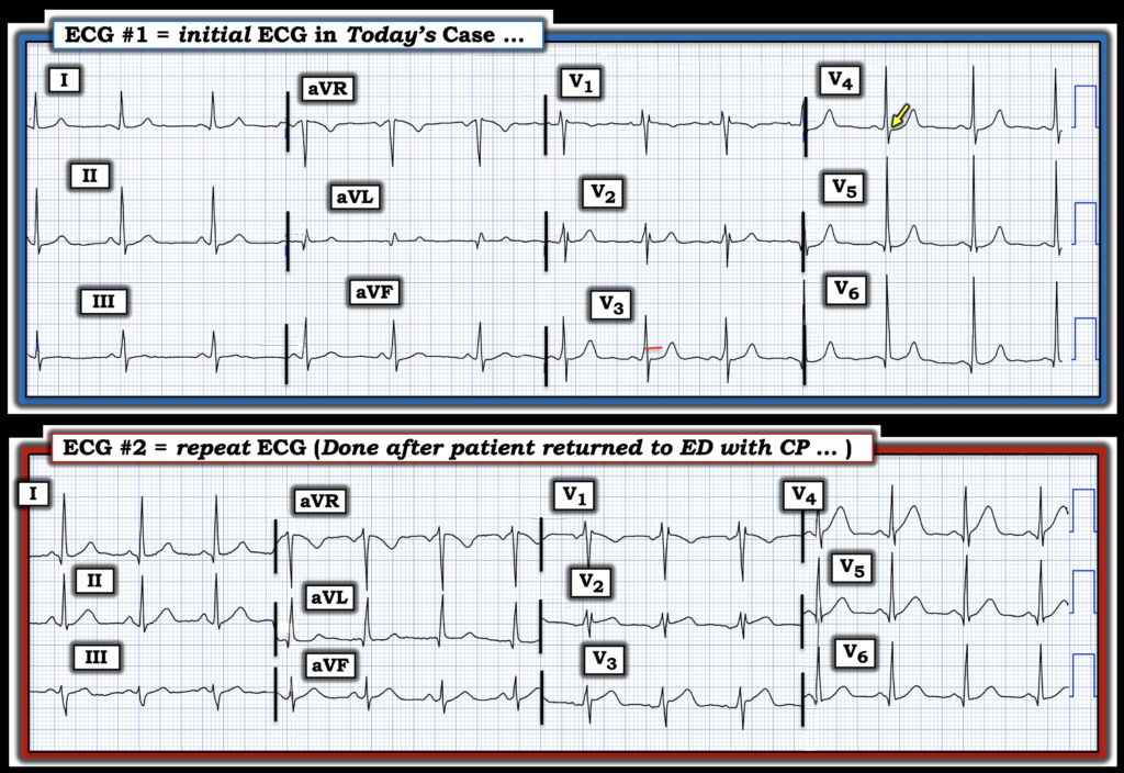
- As is often emphasized in Dr. Smith’s ECG Blog — leads V2 and V3 normally manifest slight gently upsloping ST elevation. As noted below in Figure-1 — lead V3 instead manifests ST segment straightening (RED line in lead V3).
- This is followed in neighboring lead V4 by subtle-but-real J-point ST depression (YELLOW arrow in this lead).
- Given these subtle findings in leads V3,V4 — I thought the ST-T wave appearance in neighboring lead V2 was also “off” — in that despite a nearly identical rSr’ complex in leads V1 and V2, the ST segment in lead V2 is flat and ends with an upright T wave (quite different in appearance from the ST-T wave in lead V1).
- To again emphasize — ECG #1 is non-diagnostic. But in the context that we should be looking for potentially subtle changes — I didn’t interpret this tracing as “negative”. I’d want to correlate this tracing with the presence and relative severity of patient symptoms over their time in the ED — and a repeat ECG would seem essential prior to discharging the patient.
= = =
Figure-1: Comparison between the 1st and 2nd ECGs in today’s case.
= = =
Oversight #3: How to compare serial tracings
The patient returned to the ED after being discharged because her CP worsened. The repeat ECG was “not interpreted as STEMI, and no immediate action was taken”.
- Although I was not there — by definition as we look at Figure-1, in which I’ve placed ECG #1 and ECG #2 together — lead-by-lead comparison of these 2 tracings can not possibly fail to make obvious the difference in ST-T wave appearance between these 2 tracings.
- I’ve learned the hard way that unless you look lead-by-lead when assessing serial ECGs — it is EASY to overlook “dynamic” ST-T wave changes between 2 tracings.
- Limb lead T waves are clearly more upright and “bulky” in ECG #2 (especially in leads II and aVF).
- Whereas ST segments in chest leads V3,V4 were flattened, if not slightly depressed in ECG #1 — there is now hyperacute ST elevation in leads V2,V3,V4 in ECG #2 (with ST segment straightening and much “fatter”-at-their-peak and wider-at-their-base T waves in these leads).
- Bottom Line: Regardless of whether or not sufficient ST elevation is now present in ECG #2 to qualify as a “STEMI” — these “dynamic” ST-T wave changes that are now obvious with lead-by-lead comparison in Figure-1, and which are now occurring in association with this patient’s recurrence of CP — are clear indication for immediate cardiac cath.
= = =
= = =