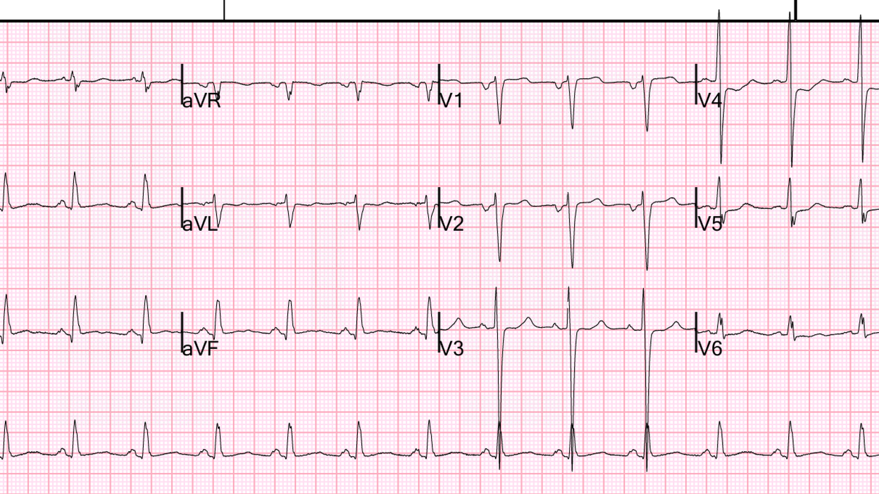
This was sent to me by an internal medicine MD who was looking back on the chart of one of his patients that he was seeing in his clinic.
This 30-something young woman with history of hypertension and obesity that is resolved (appropriate weight loss) reported experiencing right arm and upper “burning” chest pain for the past 10 days, on and off.
She has had similar symptoms in the past and was put on omeprazole and was told to undergo endoscopy. The patient was previously taking omeprazole but stopped several weeks ago on the advice of her doctor to allow for H. pylori testing; she notes that the pain has been present even prior to stopping the medication. She also reports new swelling in her left leg and experiences shortness of breath during the painful episodes of vomiting.
One month prior she was in the clinic complaining of epigastric pain, but also complained of chest pain at that time.
Here is the note:
Chest Pain and Associated Symptoms
– Experienced a severe burning chest pain episode two nights ago, starting at 9:00 PM and lasting until 4:00 AM.
– Episode was accompanied by vomiting, dehydration, and fatigue.
– Unable to retain fluids during the episode.
– Described the pain as “excruciating” and distinct from prior episodes.
– Similar episodes recur, sometimes with elevated blood pressure, especially at night when relaxing.
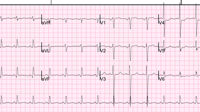
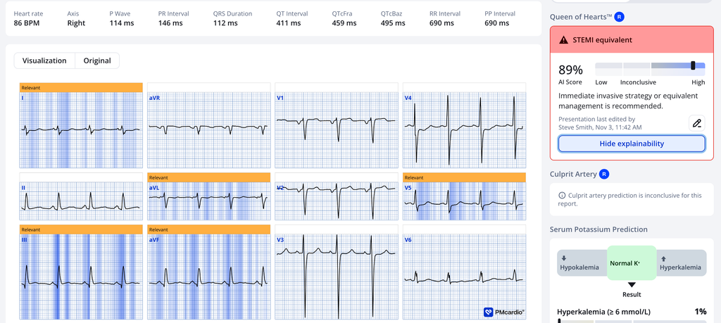
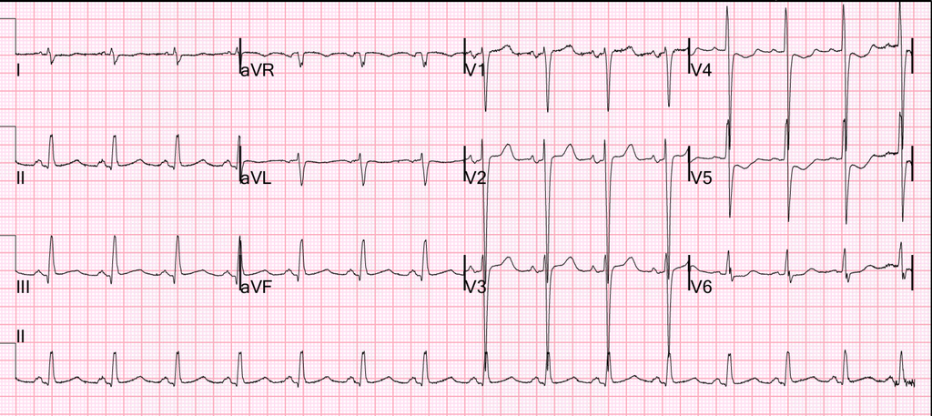
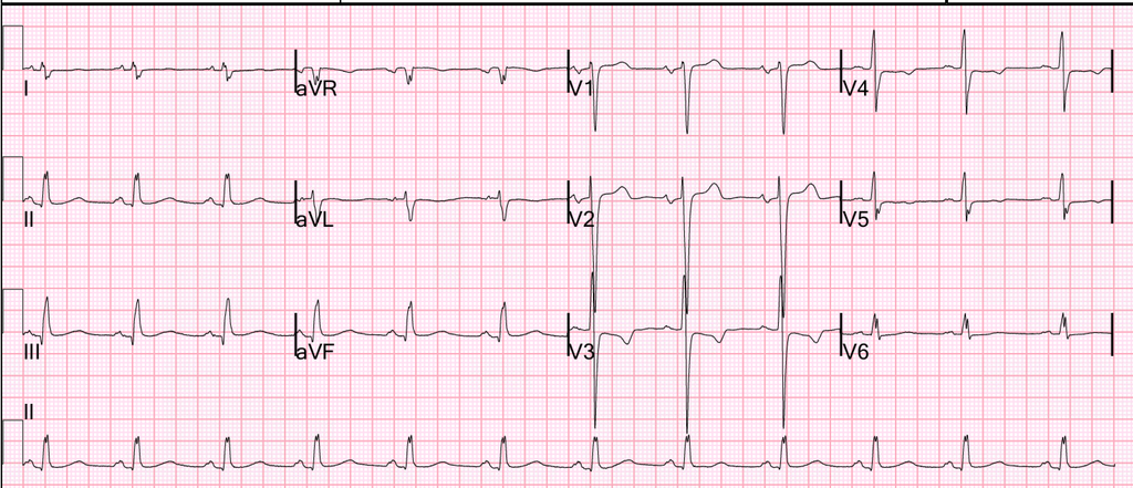
Here is the ECG recorded at that time, more than a day after the symptoms resolved:
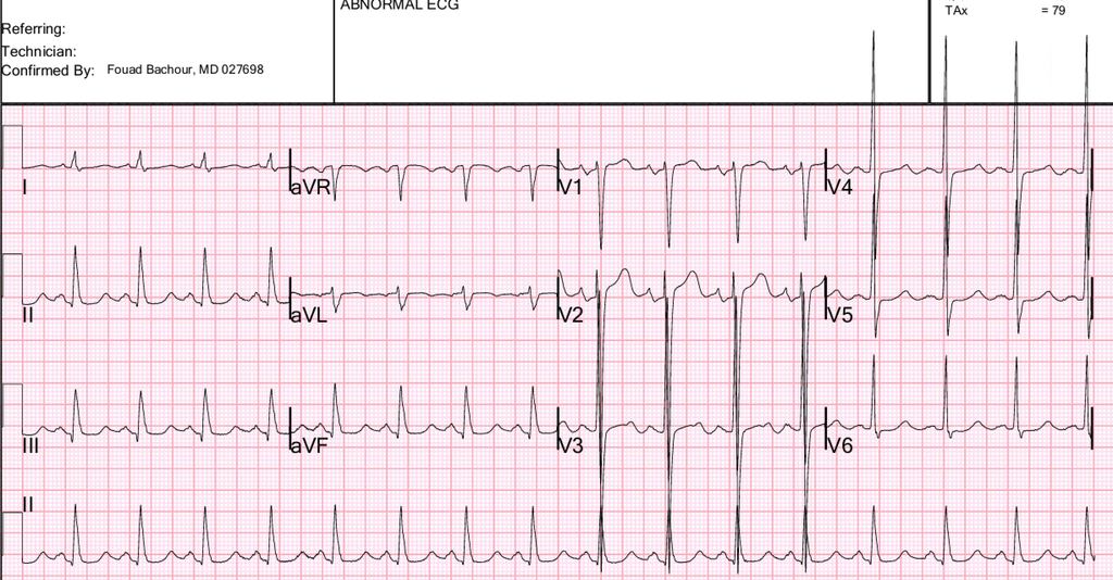
This shows LVH but without any signs of ischemia, and no significant ST depression. The PMCardio Queen of Hearts diagnosis was “No OMI” and “Left Ventricular Hypertrophy”
No troponin was measured and she was discharged to home.
ED Case continued:
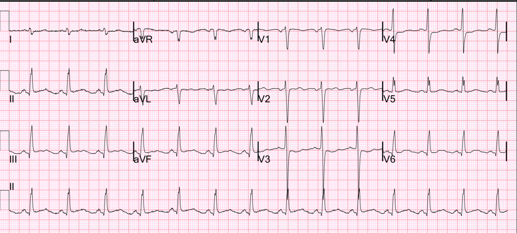
She was hypertensive with a blood pressure of 174/118. Here is her ED ECG with chest pain:
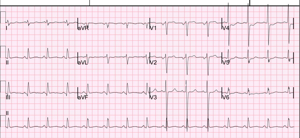
What do you think?
This is a comparison with the previous ECG from one month prior:
This compares leads III and aVL between the 2 ECGs. I hope you can see the very diagnostic difference:
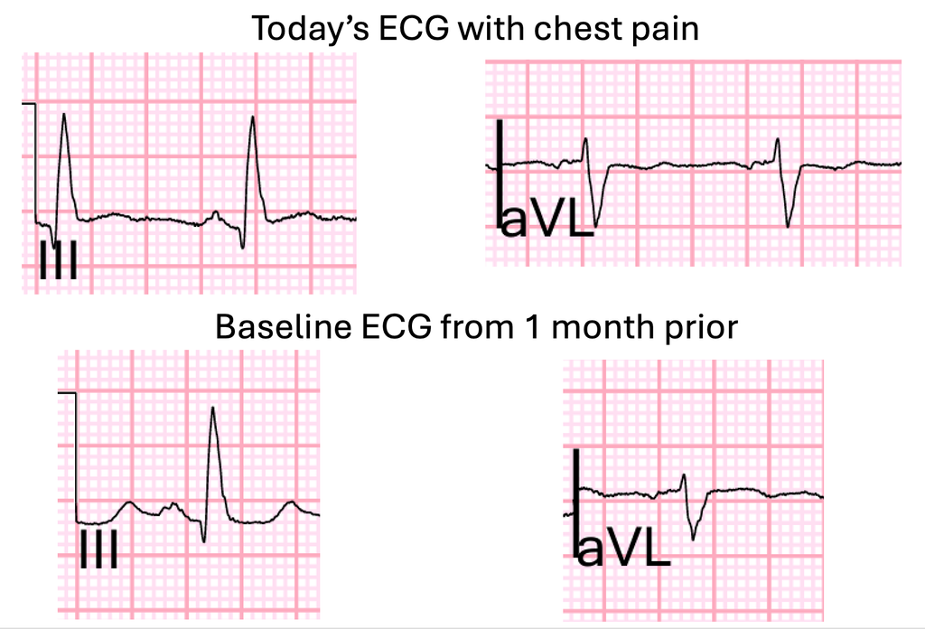
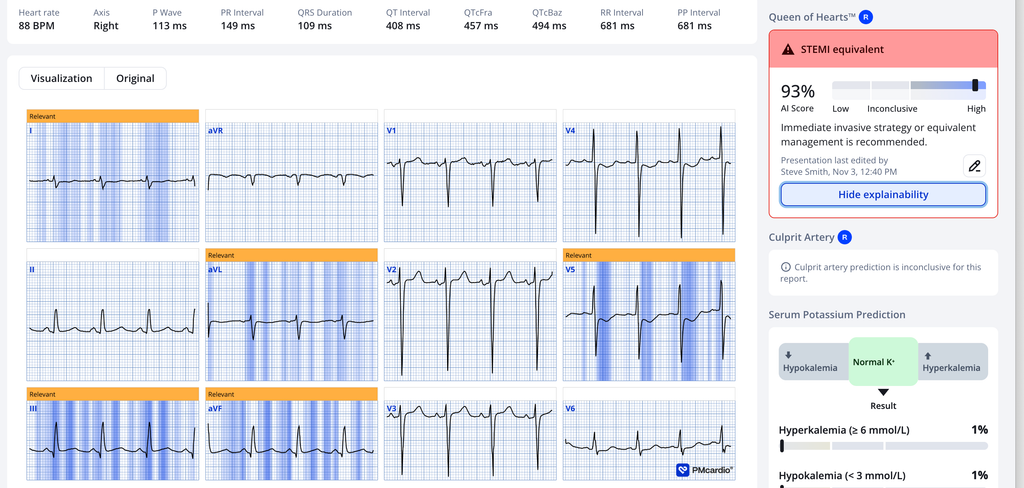
This ECG is diagnostic of inferior-posterior OMI. There is subtle STE with a straight ST segment in lead III with reciprocal ST depression and down-up T-wave in aVL. There is ST depression maximal in lead V4.
Here is the PMCardio Queen of Hearts AI Model’s interpretation:
No one noticed that there was any evidence of occlusion. “ST depression” was mentioned in the chart, but along with “no acute finding” (!!!!).
“No STEMI criteria” was documented.
A bedside ultrasound was done by an ultrasound-fellowship trained emergency physician and no wall motion abnormality was identified. There was concentric LVH noted.
Here are the ultrasound videos:
I am not an ultrasound expert, but I can see that this ultrasound has poor LV function AND at least an apical wall motion abnormality.
She reported that her chest pain had significantly improved, but was still present, after receiving a GI cocktail (Maalox and Lidocaine) in the ED.
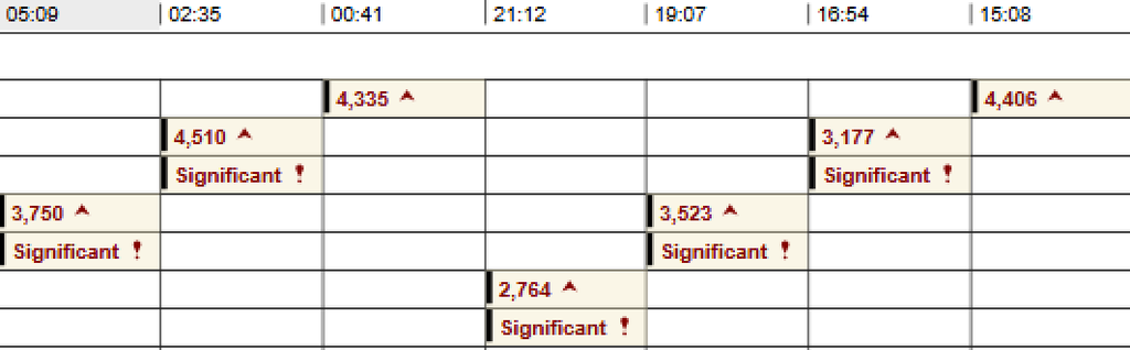
The first troponin returned at 4406 ng/L.
Now there can be no doubt about the diagnosis.
Aspirin was administered and Cardiology was consulted and provided the following recommendations:
- Treat as NSTEMI until proven otherwise.
- Initiate nitroglycerin infusion.
- Initiate heparin infusion.
- Start carvedilol 6.25 mg twice daily.
- Continue aspirin 81 mg daily.
- Start atorvastatin 40 mg daily.
- NPO after midnight.
- Maintain blood pressure with goal systolic <140 mmHg and diastolic <100 mmHg.
- Re-contact Cardiology for recurrent chest pain or significant EKG changes.
- Admit to step down unit for further management.
After a dimer returned at 770 mg/L, CT pulmonary angiogram was ordered and was negative for any acute cardiopulmonary abnormality. This was clearly unnecessary.
Repeat ECG was recorded 4 hours after the first:
The ECG remains ischemic even though the chart reports that the pain had improved.
Repeat troponin was a bit lower at 3177 ng/L. This is somewhat reaasuring, but troponin only tells you what was going on hours before, NOT what is going on at the moment.
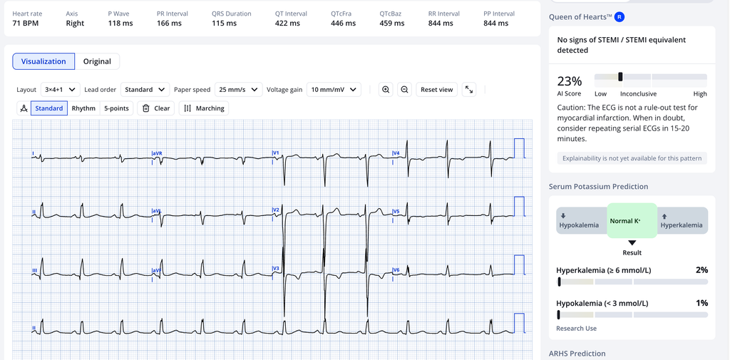
By 8 hours after arrival, the pain was gone and another epeat ECG was recorded:
This is far better, but still shows evidence of OMI:
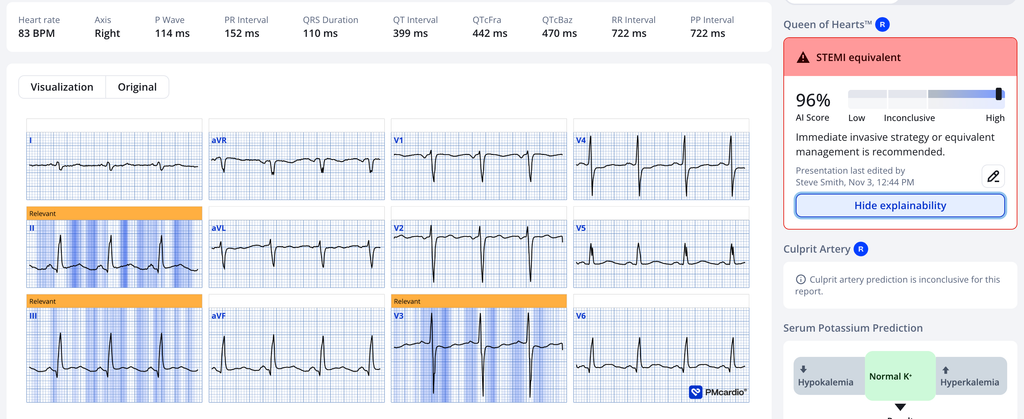
It is unclear whether the patient continued to have pain or not. No more ECGs were recorded before the next day angiogram.
Here is the formal bubble contrast echo:
The estimated left ventricular ejection fraction is 40%. Severely dilated left ventricle by volumetric measurements with mild increase in LV wall thickness. Regional wall motion abnormality- hypokinesis apical lateral, akinesis apical anterior, hypokinesis basal inferoseptum.
So this is a significantly impaired ventricle. How much of that was due to an infarction one month prior, and how much due to infarction today, is not certain.
Angiogram
Culprit for the patient’s non-ST elevation myocardial infarction is a subtotal occlusion of a large distal OM1 Suspected chronic total occlusion of a small to medium caliber RCA.
Severe, premature atherosclerosis. Culprit for the patient’s non-ST elevation myocardial infarction is a subtotal occlusion of a large distal OM1. Suspected chronic total occlusion of a small to medium caliber RCA.
Troponin profile:
2 days later
There is no cardiac follow up for this patient.
Learning points:
- GERD (gastroesophageal reflux) cannot be differentiated from myocardial ischemia without both an ECG and troponin. The pain is exactly the same.
- Acute MI cannot be ruled out with a normal ECG. Only troponin can do that
- Acute persistent occlusion is best diagnosed with the ECG, and the Queen of Hearts is now known to be 92% sensitive and specific for instantaneous occlusion (presented at TCT, San Diego, Nov 2 2026)
- Even clinics should have troponin and use it. This patient probably had a myocardial infarction that went undiagnosed at the clinic visit one month prior.
- The worst thing you can be if you have an MI is a young woman. No one will believe the ECG, the troponin, or even that you need these ordered!
- Bedside echo can be very helpful, but this was a miss. I don’t know why. Maybe because she was a young woman and the operator was just going through the motions.
- Therapeutic trials such as a “GI Cocktail” are TOTALLY UNRELIABLE as a diagnostic maneuver. By all means try to relieve symptoms by giving them, but at least 25% of ischemic chest discomfort will resolve after a GI Cocktail, because ischemic pain comes and goes!
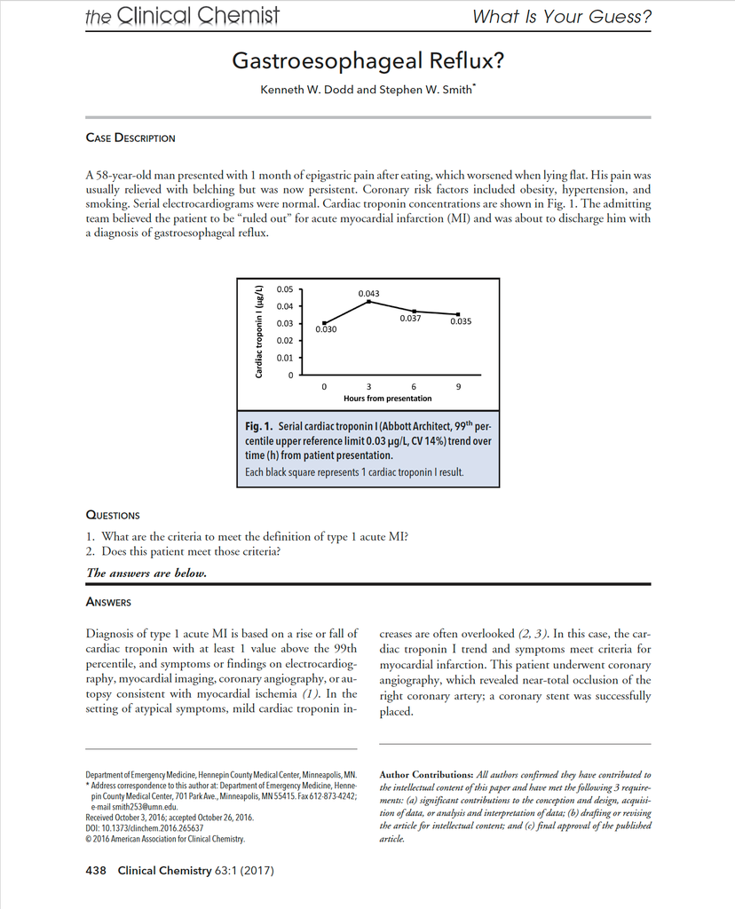
Here is an instructive case about troponin that we pubished in Clinical Chemistry 63:1 (2017). Unfortunately, I do not have the ECG and I cannot remember what it showed.
= = =
======================================
MY Comment, by KEN GRAUER, MD (11/6/2026 ):
I found today’s case challenging for a number of reasons. These include the following:
Reason #1: The History …
- The History is confusing. Although the patient is a young woman (in her 30s) in whom one might consider the risk of acute coronary disease to be less — she has known hypertension, and has had a fluctuating bodyweight.
- The patient has had overlapping symptoms — including a “burning” CP (Chest Pain) occurring “on-and-off” over what sounds like weeks, for which she has been treated with Omeprazole for a presumed GI etiology. In addition — she reported some leg swelling with shortness of breath and painful episodes of vomiting.
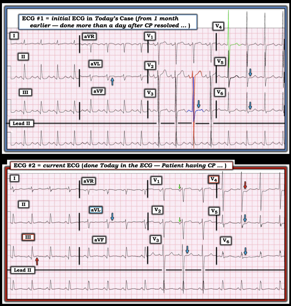
- She was seen in clinic for these symptoms a month prior to her presentation to the ED in today’s case. At that time — she described an episode of severe “burning” CP that lasted for hours, but which had resolved more than a day before her clinic visit (at which time ECG #1 that I’ve reproduced in Figure-1 below was recorded).
- ECG #1 was apparently not felt to indicate a cardiac etiology (We are not told how this ECG was interpreted — only that no Troponin was ordered and the patient was discharged home).
- Apparently, the patient’s intermittent symptoms of epigastric (GI-sounding) pain, as well as “burning” CP continued for another month — ultimately resulting in today’s ED visit. The patient was having CP at the time her ECG in the ED ( = ECG #2) was recorded.
Bottom Line: I found the history in today’s case difficult to put together. Providers were apparently misled by the long duration of a combined set of intermittent symptoms suggesting GERD, but also including recurrent “burning” CP.
- As per Dr. Smith — the symptoms of GERD can not be reliably distinguished from ACS (Acute Coronary Syndrome) on the basis of history alone.
- Although this patient’s symptoms were intermittent — they were ongoing for weeks (if not months) without a positive response to Omeprazole, and therefore without a definite diagnosis. Given a component of recurrent CP — the lesson to be learned is the need to rule out coronary disease sooner.
= = =
Reason #2: The ECGs are not “normal” …
I thought the first 2 ECGs in today’s case insightful for a number of subtle but important reasons. For clarity in Figure-1 — I’ve reproduced these first 2 tracings.
- ECG #1 — is the tracing recorded a month prior to today’s ED visit. This tracing is remarkable for sinus tachycardia (rate ~100/minute) and marked LVH. I’ve color-coded QRS complexes in the mid-chest leads to facilitate assessment of the huge voltage amplitudes rendered confusing by lead overlap (RED = an S wave >60 mm in lead V3; GREEN = a combined R+S wave >50 mm in lead V4). The magnitude of these increased QRS amplitudes tells us that this younger adult with known hypertension is already manifesting significant structural disease.
- To emphasize — marked LVH in the absence of acute ST-T wave changes is not a reason to admit the patient to the hospital. But as per Dr. Smith — a single ECG, especially in the absence of Troponin — was not enough to rule out ACS on this patient’s clinic visit a month earlier.
- KEY Point: The fact that ECG #1 manifests both sinus tachycardia and such marked LVH makes it that much more difficult to detect subtle acute changes. This is all the more true given that the patient’s CP had resolved more than a day before ECG #1 was recorded in clinic (ie, It’s that much less likely that an ECG recorded more than a day after CP resolves will still show acute changes).
- For example — the subtle ST segment coving with slight ST elevation in lead aVL of ECG #1 (BLUE arrow) is not the ST-T wave response I’d expect in a lateral lead from simple LVH.
- The same for the ST segment flattening as highlighted by BLUE arrows in the chest leads (with ST depression in lead V6).
- To Emphasize: ECG #1 is non-diagnostic. But I find it suspicious that an acute event may have occurred at the time of this patient’s episode of severe “burning” CP that lasted for hours, but which resolved more than a day before ECG #1 was recorded.
- Lesson-to-be-Learned: This patient with known hypertension clearly will have prior ECGs in her records. Prior to discharging her home from the clinic without a Troponin — the least that should have been done would be to access a prior ECG for comparison to see if the above noted subtle ST-T wave changes might be new.
= = =
ECG #2: Done during Chest Pain …
A number of important findings were missed on ECG #2 — which is the tracing recorded during CP, at the time the patient presented to the ED in today’s case.
- As per Dr. Smith — ECG #2 is diagnostic of acute infero-postero OMI until proven otherwise. Although by itself — the amount of ST elevation in lead III and the amount of mirror-image opposite ST depression in lead aVL is subtle — this is clearly a new change compared to the scooped ST segment shape in lead III and the coved ST segment shape in lead aVL that was present in ECG #1.
- Lesson-to-be-Learned: Isn’t it so much easier to appreciate differences between serial ECGs when you assess both tracings placed side-by-side? (as in Figure-1).
- It should also be clear from comparison between ECG #1 and ECG #2 — that there is more chest lead ST depression in ECG #2, which is especially marked in lead V4 — and which suggests recent posterior OMI (RED arrow in lead in lead V4).
- NOTE: The P waves in leads V1 and V2 in ECG #2 are all negative. This is also a new change since ECG #1 (The P wave in leads V1,V2 in ECG #1 was biphasic with a smaller negative component). This finding suggests too-high lead placement of lead V1,V2 in ECG #2 — which is a relevant finding given how suspicious we are of acute posterior OMI (See the Addendum to My Comment in the March 6, 2026 post — for more on lead V1,V2 misplacement). Repeating ECG #2 after correcting the erroneous lead placement would probably have shown some ST flattening/depression in leads V1,V2 that would further support our strong suspicion of posterior OMI.
Finally — Note dramatic change in the pattern of QRS morphology between ECG #1 and ECG #2.
- As noted above — We already know that the lead V1,V2 electrodes have been misplaced.
- The fact that the patient is a woman in whom QRS voltage is markedly less in the chest leads in ECG #2 compared to ECG #1 — is probably the result of an additional difference in precordial electrode lead placement.
- Malposition of chest lead electrodes is common in general practice (Rehman and Rehman — Cureus 12(7):e9040, 2020). Because of placement issues with respect to breast tissue — optimal chest lead electrode placement is especially problematic in women — which leads to inconsistent, and all-too-often, technician dependent variations (Kligfield et al — AHA/ACC/HRS Scientific Statement, 2007).
- In particular — the abrupt drop-off in R wave amplitude in the lateral chest leads of ECG #2 may well be related to lateral chest lead electrodes being placed on top of (rather than under) breast tissue.
- There is no universal consensus on whether to place chest lead electrodes V4,V5,V6 on top of or underneath breast tissue (Rautaharju et al — J Electrocardiol 31(1):17-29, 1998). What is important — is consistency, as much as this is possible. This is because when different technicians use different landmarks and different techniques when recording ECGs on women — potentially marked differences in both QRS and ST-T wave morphology may result.
- How much a difference in recording technique influenced the different QRST appearance in the chest leads for the 2 tracings in Figure-1 is uncertain.
- My Thoughts: I favor Anatomic/On-Breast Approach — in which chest lead electrodes are placed on top of breast tissue over the true anatomical intercoastal landmarks. This is because data suggests that breast tissue exerts no more than a modest effect on QRS amplitudes — whereas placing the electrodes underneath breast tissue may significantly displace the leads downward, with resultant distortion of QRST morphology (Derkenne et al — BMJ Case Rep, 2017 ).
- Other sources favor the Practical/Under-Breast Approach — in which the V4,V5,V6 electrodes are placed in the inframammary fold (under the breast). This may be helpful when body habitus renders it difficult to accurately palpate the 4th and 5th intercoastal spaces — with placement of the leads under the fold providing a readily identifiable firm surface for placement.
- Lesson-to-be-Learned: Consideration of female anatomy may prove important when comparing serial tracings, especially if significant differences in QRST morphology are noted between the 2 tracings being compared (as should be evident in Figure-1).
= = =
Figure-2: I’ve reproduced and labeled the first 2 ECGs in today’s case.
= = =
= = =