This 54 year old patient with a history of kidney transplant and poor transplant function — had been vomiting all day, when at 10 PM he developed severe substernal crushing chest pain.
- He presented to the Emergency Department with a blood pressure of 111/66 and a pulse of 117.
= = =
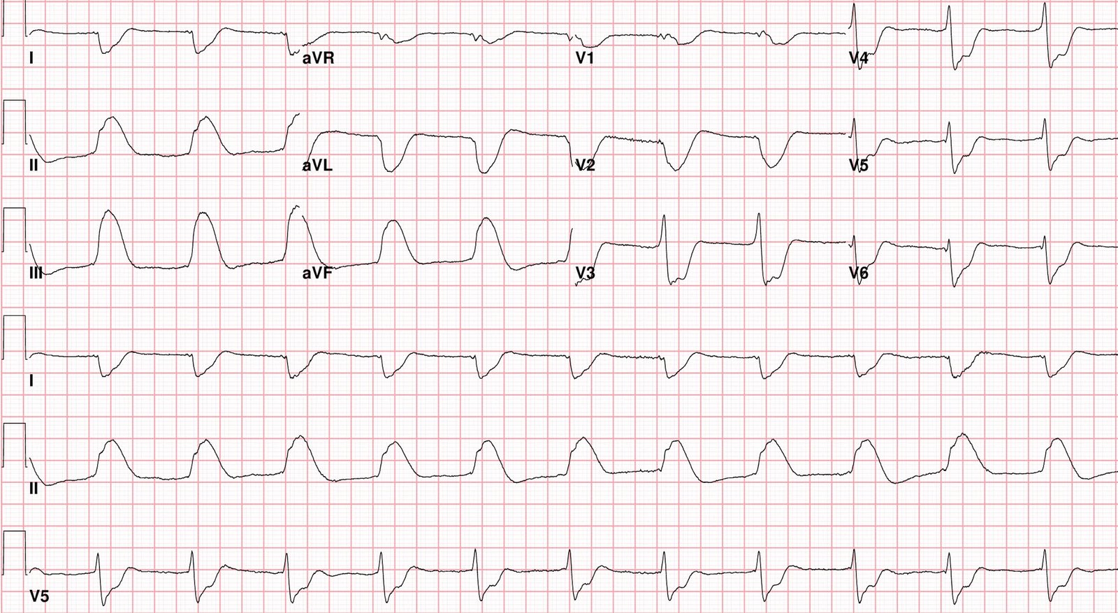
He had this ECG recorded.
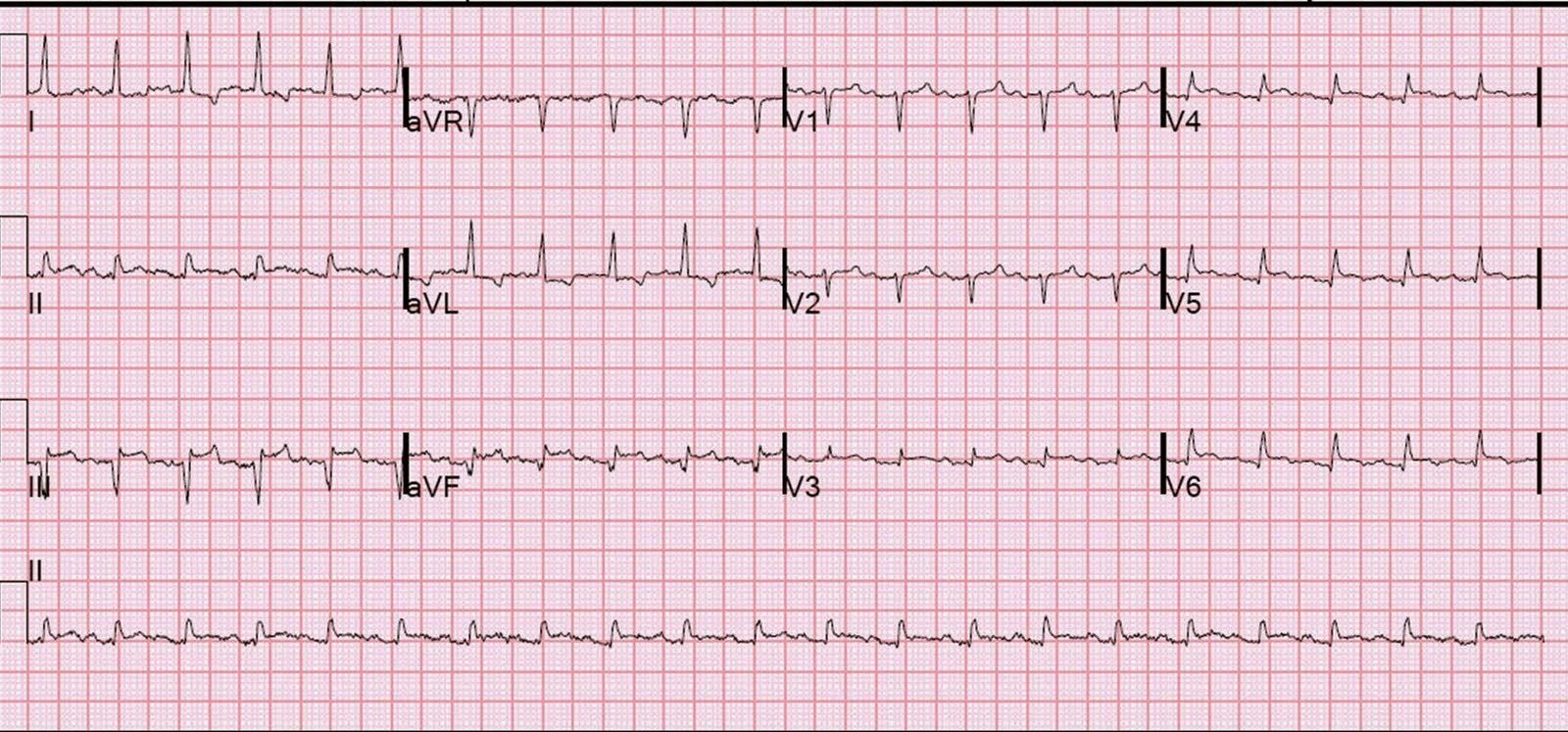
- He was rushed by residents into our critical care room with a diagnosis of STEMI, and they handed me this ECG:
= = =
There is sinus tachycardia with ST elevation in II, III, and aVF, as well as in leads V3-V6. There is reciprocal ST depression in I and aVL. At first glance, it seems the patient is having a STEMI.
But, remember, we do not evaluate and treat ECGs; we evaluate and treat patients. Even if this ECG is the first thing one sees (as it was for me) — one should stop and think: “This is an unusual STEMI.” Why?
ACS and STEMI generally do not cause tachycardia unless there is cardiogenic shock. Are the lungs clear? Is the patient cool and pale? Then ACS (STEMI) might be primary; this might be cardiogenic shock.
More often, tachycardia with ST segment abnormalities (elevation or depression) — is due to an underlying illness (PE, sepsis, hemorrhage, dehydration, hypoxia, respiratory failure, etc.). One must clearly rule out these processes before jumping on the ACS diagnosis.
Furthermore, notice the well-formed Q-waves in the inferior leads. These must raise suspicion of old MI with persistent ST elevation.
One very useful adjunct is ultrasound:
- Echo of his heart can distinguish aneurysm from acute MI by the presence of diastolic dyskinesis, but it cannot distinguish demand ischemia from ACS.
- In today’s case — bedside echo did not reveal any wall motion abnormality. Instead,there was hyperdynamic function, which is not consistent with cardiogenic shock, but rather with sepsis or volume deficit.
Large volume fluid resuscitation was undertaken.
- The K returned at 6.9 mEq/L. The HCO3 was 8. Cr was 13.4. Even after 3 liters of fluid, his CVP was very low.
Troponins peaked at 0.275 ng/ml.
An angiogram showed no acute coronary lesions. The patient was suffering from severe dehydration, possibly with sepsis.
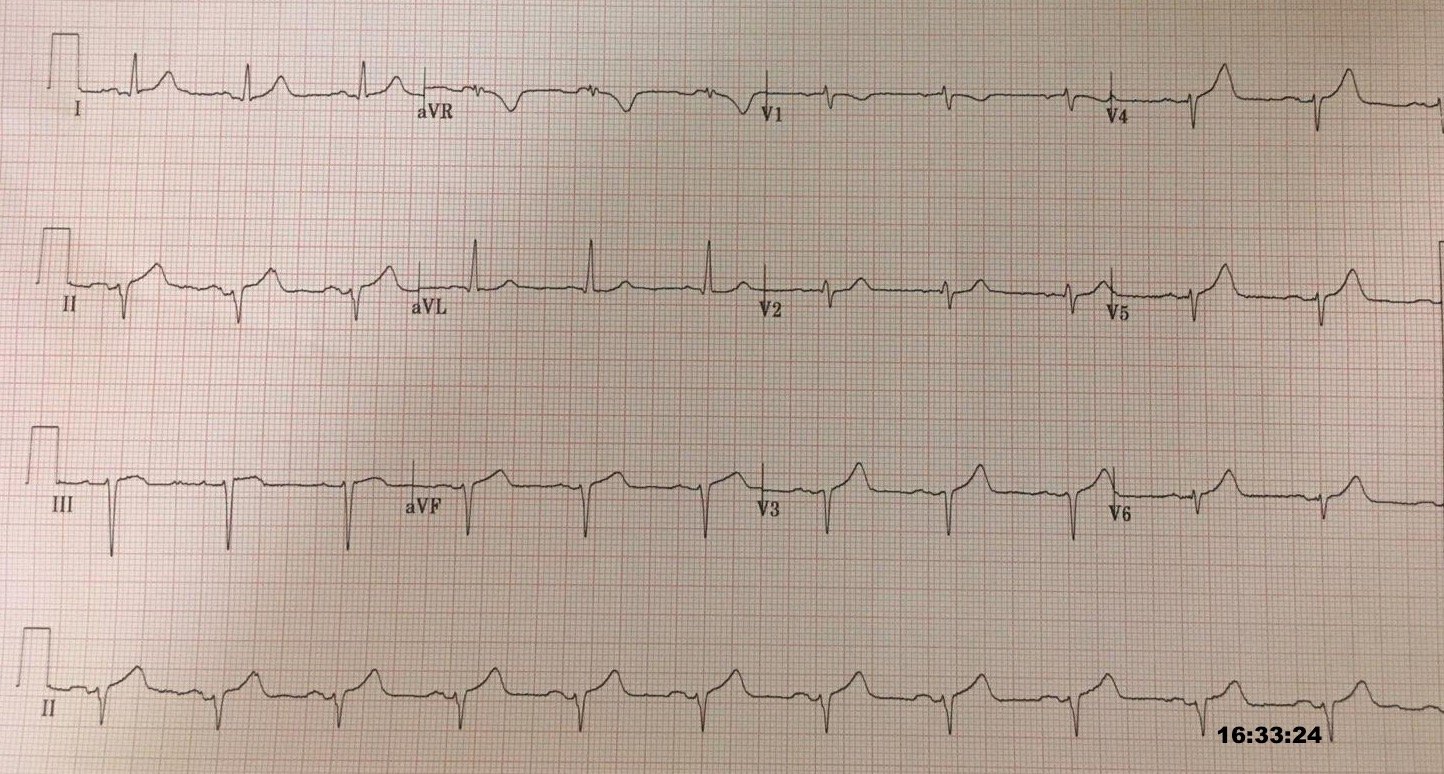
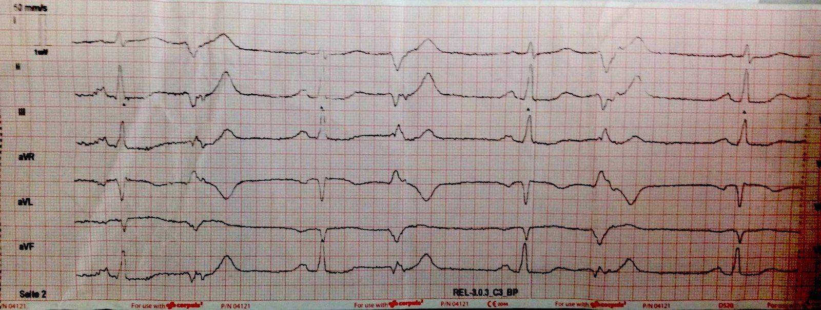
After stabilization, previous ECGs and an old echocardiogram were found:
- The prior ECGs demonstrated an old inferior MI, with persistent ST elevation (LV aneurysm morphology).
- The previous Echo showed diastolic dyskinesis.
= = =
= = =