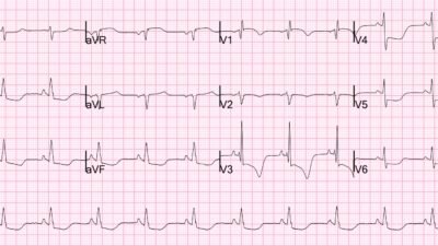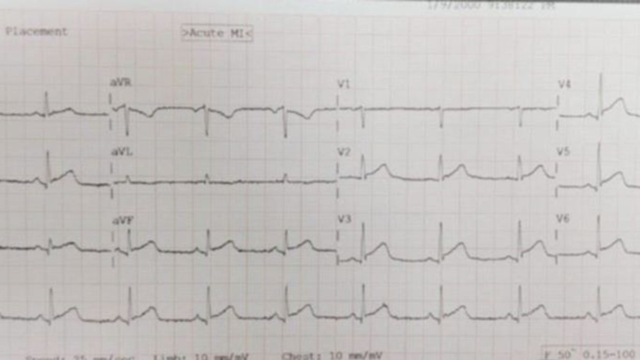
Also, listen to young doctors. They often know much more than the experienced ones.
This was sent by Ahmed Adel.
Hello, Dr. Smith. I hope you’re doing well. I’m a 2nd-year internal medicine board resident from Iraq. I’ve been following your work over the past few years, and I truly appreciate all that you’ve done. I wanted to share this interesting case with you & would greatly value any insights you might have whenever you have the time.
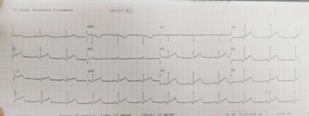
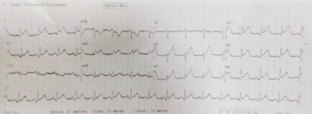
This ECG belongs to a 55-year-old man with a negative past medical history presenting with chest pain.
What do you think?
Smith: This ECG is diagnostic of LAD OMI. There is ST elevation in V2-4 and beyond. Is it normal variant? There is Terminal QRS distortion (TQRSD), which rules out normal variant. Therefore, it is due to acute LAD Occlusion until proven otherwise. There is more discussion below. TQRSD is defined in the setting of some amount of ST Elevation in V2-V4, with absence of an S-wave in either V2 or V3, UNLESS there is a prominent J-wave.
Here there is absence of an S-wave in BOTH V2 and V3, without a J-wave.
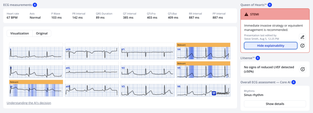
The Queen’s numerical output was 0.81, which is about 98% specific for OMI. Interesting that she does not highlight the terminal QRS distortion, except in leads V4-5! It may be true that TQRSD in V4-5 is very significant, and may also be significant in inferior leads. However, we have never studied it. The Queen has learned from tens of thousands of troponin-positive and millions of troponin negative ECGs (and she can remember, unlike we fallible humans!) so she might know better than anyone.
Case continued:
“The senior cardiologist diagnosed this as pericarditis & wanted to give the patient indomethacin & colchicine. But my colleague (who initially recieved this patient in the ED) wasn’t convinced & called me for a second opinion.”
My (Ahmed) opinion was: “lead II is entirely slanted (wandering baseline) giving the false appearance of PR depression & Spodick sign. V3 shows terminal QRS distortion (TQRSD), which is a HUGE RED FLAG for a LAD occlusion. I think it would be wise to withhold the NSAID for now and get a repeat ECG & trops before calling this a pericarditis”. About 20 min later, my colleague texted me that the patient went into VFib & was shocked back into sinus rhythm… twice!”
This ECG was recorded after defibrillation.
Now the ECG is even MORE diagnostic.
This patient was lucky to go into VF. Had he not, he might have sat there in the room for hours while myocardium was dying.
This is frequently the case. Here is another one:
Sometimes a patient is fortunate to have a cardiac arrest
See what happens when hyperacute T-waves are missed. And can you be lucky to have a cardiac arrest?
Ahmed asked this:
Would you have activated the cath lab based on clinical suspicion + the initial ECG (which only had TQRSD in V3 as the only solid clue for OMI… that I could find)? • Were there any other “red flags” in the initial ECG that were missed? • Do you know of any literature on the value of TQRSD in differentiating LADO from pericarditis? I only know of its use in LADO vs BER.
My answer: Forget pericarditis! Pericarditis is rare. I do not know of any literature, but if there is TQRSD then it is OMI until proven otherwise. Pericarditis is just a way of trying to deny that a patient is having an OMI.
= = =
==================================
MY Comment, by KEN GRAUER, MD (8/6/2026):
Today’s case once again raises the question, “Acute Pericarditis or acute OMI?”. I view the answer to this question as being comprised of 2 parts:
- Part #1: Does the initial ECG look more like acute pericarditis or like an acute OMI?
- Part #2: Taking into account the answer to Part #1 — Why is acute pericarditis unlikely?
= = =
Today’s initial ECG …
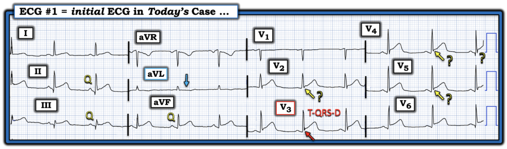
For clarity in Figure-1 — I’ve labeled today’s initial ECG. I interpreted this ECG in light of the brief history we were given = a 55-year old man with a negative past medical history, who presented with CP (Chest Pain).
- The rhythm is sinus at ~70/minute. Intervals (PR-QRS-QTc) and the axis are normal. There is no chamber enlargement.
Regarding Q–R–S–T Wave Changes:
- Q Waves: Potentially significant Q waves are present in each of the inferior leads.
- R Wave Progression: There is early transition (with a predominant R wave already present by lead V2).
- ST-T Wave Changes: There is J-point ST elevation in multiple leads (ie, leads I,II,III; aVF; and V2-thru-V6). The ST segment takeoff is straightened in most of these leads.
- The ST segment is flattened in leads aVL and V1 — with a hint of ST depression in lead aVL (BLUE arrow in this lead).
= = =
Impression: Acute Pericarditis vs Acute OMI?
Today’s initial ECG does manifest a number of features that could be consistent with acute pericarditis. These include:
- ST elevation in multiple leads (as noted above — ST elevation is present in 9/12 leads!).
- There is no loss of R wave forces in the chest leads (with a predominant R wave present already by lead V2).
- If one uses the PR segment as the ST segment baseline — then the ST/T wave ratio in lead V6 appears to be greater than 0.25 (See My Comment at the bottom of the page in the September 28, 2025 post for “My Take” on this an other ECG findings in acute pericarditis).
ECG features more consistent with acute OMI include the following:
- The ST elevation in multiple leads looks hyperacute.
- Potentially significant Q waves are present in each of the inferior leads (with ST elevation seen in each of these leads). Given a frontal plane axis that is not vertical — it would be unusual to see “normal septal Q waves” in these leads (and depth of the Q in lead III is nearly half the size of the R wave in this lead).
- Although difficult to assess given the tiny QRST amplitude in lead aVL — I interpreted the flattened and ever-so-slightly depressed ST segment in this lead as suggestive of reciprocal ST depression (which should not be present with acute pericarditis).
- If one uses the T-P segment as the ST segment baseline — then the ST/T wave ratio appears to be less than 0.25 (which is against pericarditis).
- The ST-T wave in lead II resembles that seen in lead III more than that seen in lead I (which is more commonly seen with acute OMI).
- Significant PR segment depression is lacking. (Generalized PR depression with PR elevation in lead aVR is a “soft” criterion sometimes cited in favor of acute pericarditis — albeit specificity of this criterion is poor).
- KEY Point: There is T-QRS-D (Terminal QRS Distortion)!
= = =
Figure-1: I’ve labeled today’s initial ECG. (To improve visualization — I’ve digitized the original ECG using PMcardio).
= = =
Regarding T–QRS–D:
As emphasized in Dr. Smith’s above discussion, the importance of recognizing T-QRS-D — is that IF this unique ECG finding is present, it essentially rules out repolarization variants and rules out acute pericarditis — and — it essentially rules in acute OMI (For illustration and review of T-QRS-D — Check out My Comment at the bottom of the page in the May 8, 2025 and January 19, 2026 posts).
- T-QRS-D — is defined as the absence of both a J-wave and an S-wave in either lead V2 or lead V3 (and probably also in lead V4). Although simple to define — this finding may be subtle!
- A J-wave may manifest as either a notching or slurring that occurs where the end of the QRS joins the beginning of the ST segment (ie, the J-point joins the S of the QRS to the beginning of the ST segment).
To Emphasize: T-QRS-D is not a common finding with acute OMIs. But because of the diagnostic specificity of T-QRS-D when it is found — I carefully considered leads V2-thru-V5 in Figure-1 for the possibility of this finding.
- T-QRS-D is clearly present in each of the 3 beats seen in lead V3!
- It appears to also be present for the middle complex in both leads V2 and V4 — but it is not clearly present for the other 2 beats in these leads.
- It appears to be present in each of the 3 beats in lead V5 — but data is lacking regarding specificity of T-QRS-D in lead V5.
= = =
Part #2 The “Bottom Line” regarding this ECG:
I could not rule out the possibility of acute pericarditis solely on the basis of the ECG in Figure-1 — because there is ST elevation in multiple leads. That said:
- The reason Dr. Smith’s statement that, “You diagnose acute Pericarditis at your Peril”always echoes in my mind — is that in practice, acute Pericarditis is a rare diagnosis (especially when a previously healthy middle-aged man presents with new CP — in whom an acute OMI is much, much more commonly seen). Therefore — We need to assume acute OMI until proven otherwise.
- The finding of T-QRS-D in lead V3 strongly supports the diagnosis of acute OMI until proven otherwise.
- The consequences of missing a diagnosis of acute pericarditis are not life-threatening. In contrast — the consequences of missing the acute OMI that today’s patient presented with were almost lethal!
- Fortunately — today’s patient was successfully resuscitated from a cardiac arrest, that at least served to call attention to his acute OMI in need of prompt cath and PCI.
= = =
Additional Points:
- Even if there would have been convincing clinical suspicion of acute pericarditis in today’s case — We need to remember that the onus of proof is on the clinician to prove that the patient in front of them does not have an acute OMI. This is done by repeating the ECG soon (ideally within 10-20 minutes of the initial ECG) — by performing Echo at the bedside (looking for a localized wall motion abnormality or pericardial effusion that might be diagnostic) — and by serial Troponins.
- It’s hard to tell from the history that we were provided with — IF a pericardial friction rub was carefully listened for? Hearing a rub (as well as finding a significant pericardial effusion on Echo) are about the only ways to clearly diagnose pericarditis at the bedside.
- Given the clinical significance of recognizing what is (and what is not) T-QRS-D — it may prove insightful to check out My Comment at the bottom of the page in the January 30, 2026 post for an example of when the diagnosis is not acute OMI (as well as for an excellent illustration regarding the subtleties of determining whether T-QRS-D is or is not present).
= = =
= = =