This case was sent to me by Ahmed Marai, who is a medical student. Ahmed received it from Omer Mohammed, a fifth-year medical student from Iraq. Omer started reading Dr. Smith’s ECG blog only 2 months ago.
Omer was in the hospital when he came across this case.
A 70-year-old man presented to the emergency department at time zero with persistent retrosternal chest pain of approximately 6 hours’ duration, accompanied by shortness of breath. He was an active cigarette smoker. His past medical history was significant for hypertension, ischemic heart disease, and heart failure. He had experienced a similar episode of chest pain approximately 2 months earlier.
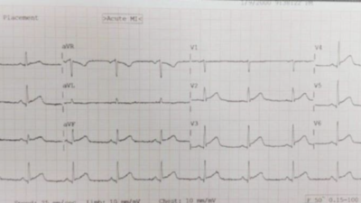
An ECG was obtained immediately upon arrival.
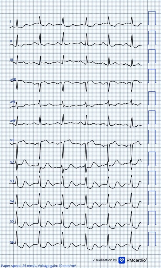
What do you think?
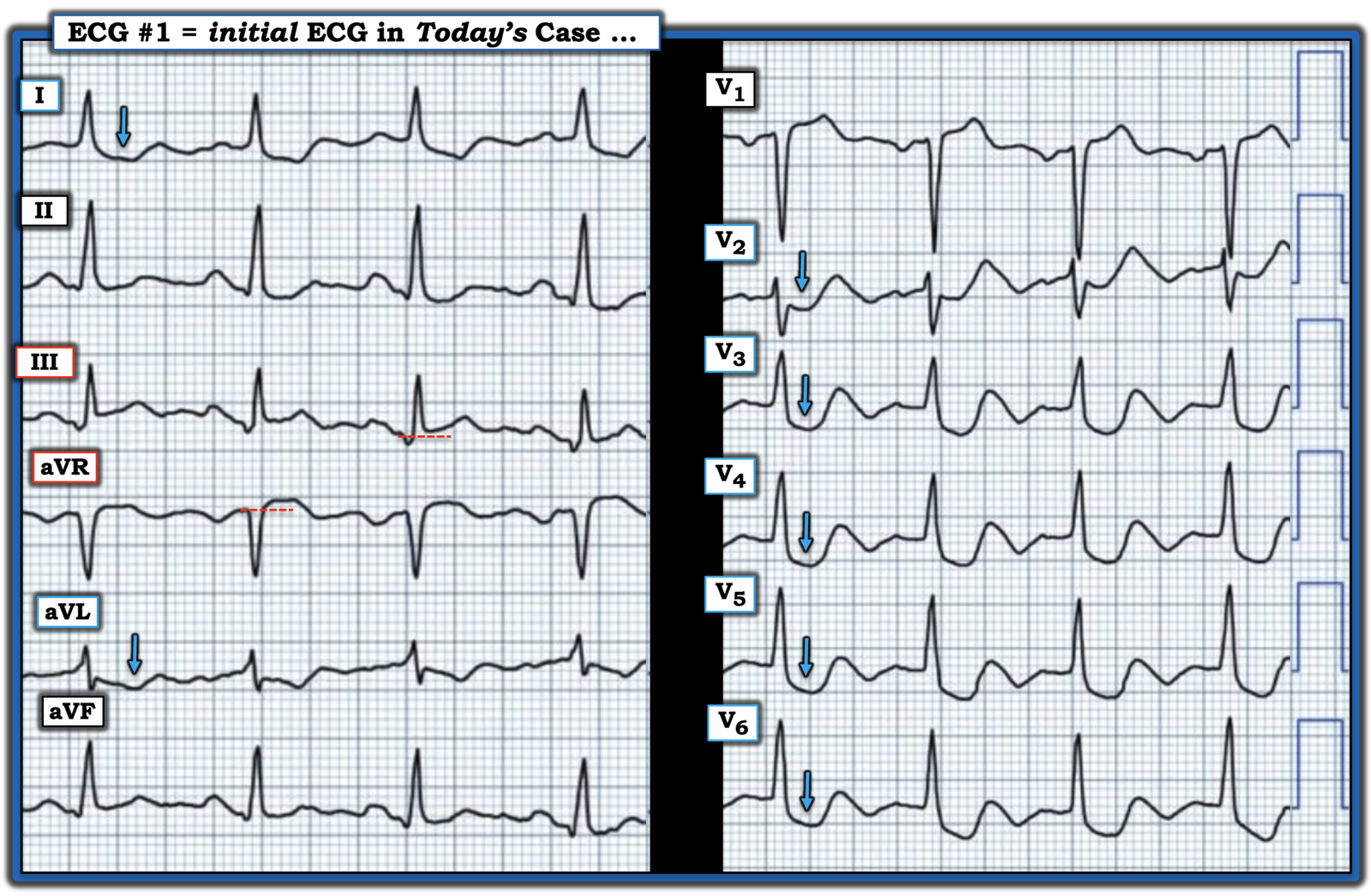
Omer saw it later and thought it was Aslanger’s pattern with posterior OMI. I agree. It is diagnostic of OMI (acute coronary occlusion).
It is a hybrid of:
1) Aslanger’s pattern (which is inferior OMI, with STE in single limb lead III, simultaneous with subendocardial ischemia (“SEI”, profound ST depression in V5-6) and
2) Posterior OMI (profound ST depression in V2).
So it is a combination of Aslanger’s with posterior OMI.
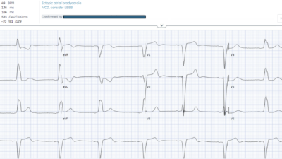
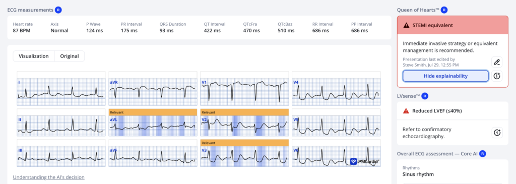
The PMCardio Queen of Hearts AI Model sees the inferior/posterior component, but our other Queen AI Model which diagnosis SEI only gave a score of 24.6% for SEI, and the Queen does not highlight V5-6:
The first high-sensitivity serum troponin I result became available 69 minutes later and was 0.030 ng/mL.
Comment on the troponin: A URL of 0.300 ng/mL is typical of 2nd or 3rd generation troponins; 4th generation troponins are the last to use ng/mL units. 5th generation are high sensitivity and report in whole numbers with units of ng/L, or sometimes pg/mL; pg = picograms.)
The patient was managed as under the totally worthless diagnosis of “NSTEMI.“
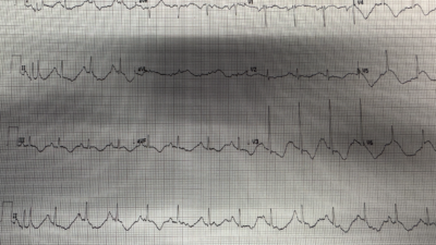
Another ECG was recorded at 120 minutes, while the patient was still experiencing chest pain.
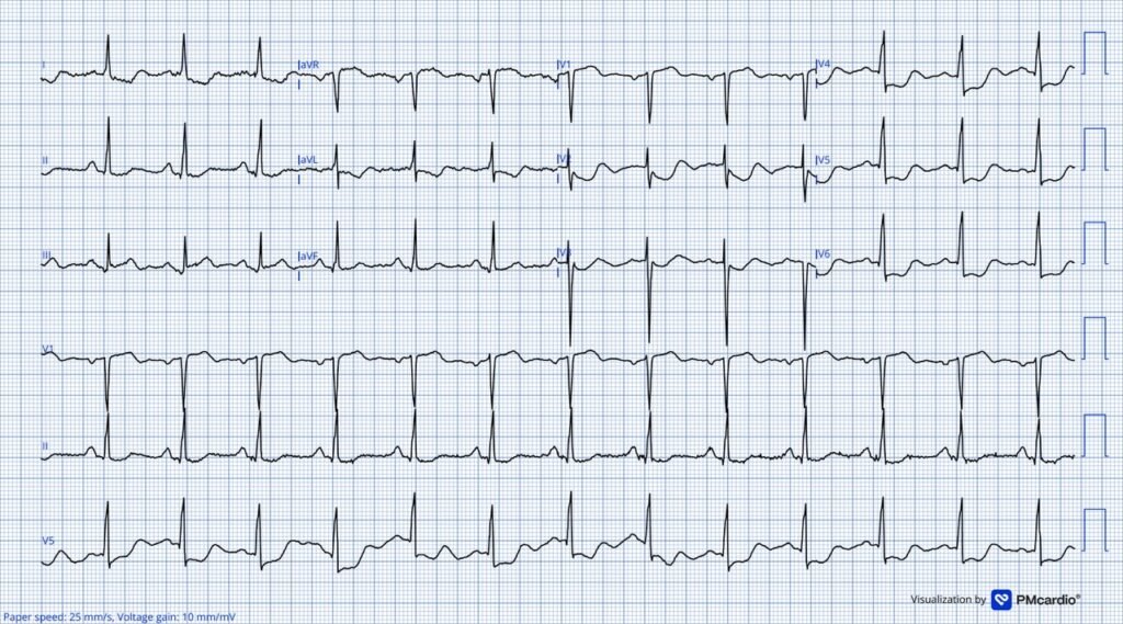
Shows the same alarming pattern
4.5 hours after arrival: Ahmed Marai: The second high-sensitivity serum troponin I result came back at 717 ng/L (apparently this one was really a high sensitivity troponin?).
5 hours 20 minutes after arrival, he was transferred to the coronary care unit under the cardiology service with a diagnosis of NSTEMI. Because the pain was severe, he received Tramadol as an analgesic.
Smith: How many times have we seen this error? Treating ischemic pain with analgesics rather than reperfusion is associated with high mortality because it hides the ischemia!! Morphine or other analgesics should only be used after the patient is committed to emergent cath lab.
See this post: What happens when you give morphine for chest pain in ACS? And what is pseudo-normalization of T-waves?
The pain decreased slightly, however, it did not resolve completely. The patient was given an appointment for delayed catheterization 2 days later.
This was when Omer came across the patient for the first time while he was training in the coronary care unit. He immediately recognized that it might be Aslanger’s pattern with posterior OMI. He went to the patient’s physician and told him that the patient might need immediate reperfusion. It turned out that the physician did not know about OMI. Omer started to explain the ECG findings to him and showed him some similar cases in his phone, but the physician was not convinced. He said, “This is just a Non-STEMI.”
Omer went to another physician, but he found no one who would listen to him. No one took him seriously because he was a student, he was not a decision maker.
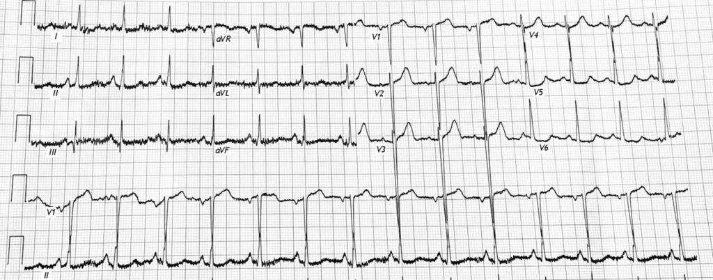
At 8:40 pm that day, another ECG was recorded. The patient still had chest pain.
Next day, at 5:45 pm, the third high-sensitivity serum troponin I result came back, and was >15,000 ng/L (15,000 ng/L is the maximum value measured by the hospital laboratory). The patient that day continued having chest pain.
The patient remained in the coronary care unit, waiting for his cath lab appointment the next day, but he was not able to reach the cath table because he died on the morning of that day, just before he was taken to the cath lab.
Learning points
- NSTEMI is a worthless diagnosis. NSTEMI diagnosis kills. Millimeters kill
- Students can be better at EKGs even than cardiologists. One need not be a cardiologist, or emergency physician, or even a doctor, to be expert at reading for OMI on the ECG. One needs to work hard, pay attention, and ignore authority. Then, when a case presents itself, one needs to dig deep into the case, and realize that those who should know what the ECG findings are, and should know how best to manage “NSTEMI,” are often frighteningly ignorant.
- Often, students and paramedics and EKG techs are far better than their MD counterparts.
- See here cases sent by Hans Helseth (who was an EKG tech at the time and has now just finished his first year of med school) and also Jack Cahill, who is a med student.
- Use the PMCardio Queen of Hearts AI ECG Model to diagnose OMI (acute coronary occlusion)
- Morphine:
- Never give opiate pain relief unless you are committed to the cath lab!! You will obscure the symptoms and think that your medical management is working to relieve ischemia, when it is only relieving pain!!
Opiates are associated with worse outcomes in Myocardial Infarction.
See this case: A man his 50s with chest pain. What happens when you treat with morphine rather than with reperfusion?
—-See this study showing an association between morphine and mortality in ACS:
Use of Morphine in ACS is independently associated with mortality, at odds ratio of 1.4. Meine TJ, Roe M, Chen A, Patel M, Washam J, Ohman E, Peacock W, Pollack C, Gibler W, Peterson E. Association of intravenous morphine use and outcomes in acute coronary syndromes: Results from the CRUSADE Quality Improvement Initiative. Am Heart J. 2005;149:1043–1049.
And an abstract that we wrote:
—-Bracey, A. Meyers HP. Smith SW. Wei L. Singer DD. Singer A. Association between opioid analgesia and delays to cardiac catheterization of patients with occlusion Myocardial Infarctions. Academic Emergency Medicine 27(S1): S220; May 2020. Abstract 556.
Main result: STEMI(-) OMI Patients
65 (23.9%) patients were found to have STEMI(-) occlusion myocardial infarction (OMI) at the time of cardiac catheterization. The 45 patients with STEMI(-) OMI without pre-cath opioids had a door-to-balloon time of 75 minutes, vs. 684 minutes for the 25 STEMI(-) OMI with pre-cath opioids.
High Risk ACS guidelines are only followed in 6% of patiients:
Lupu L, Taha L, Banai A, Shmueli H, Borohovitz A, Matetzky S, Gabarin M, Shuvy M, Beigel R, Orvin K, Minha S ’ar, Shacham Y, Banai S, Glikson M, Asher E. Immediate and early percutaneous coronary intervention in very high-risk and high-risk non-ST segment elevation myocardial infarction patients. Clin Cardiol [Internet]. 2022;Available from: https://onlinelibrary.wiley.com/doi/10.1002/clc.23781
= = =
==================================
MY Comment, by KEN GRAUER, MD (8/2/2026):
It is very sad when a patient has to die before a knowledgeable student is listened to.
- As one who taught cardiology concepts to medical students, residents and fellow physicians in practice during my 3 decades in academic medicine — what bothered me most were encounters with those who thought they knew all the answers, and who therefore refused to consider alternative possibilities to their own convictions. These individuals are dangerous clinicians.
- These concerns are especially relevant at the present time in the domain of the patient who presents with new CP (Chest Pain), and whose initial ECG is not clearly positive for STEMI criteria.
- In Dr. Smith’s ECG Blog — We’ve shown countless cases of patients with acute OMI who fail to satisfy STEMI criteria on ECG — but who eventually undergo cardiac cath that confirms acute coronary occlusion that was evident hours (and sometimes days) earlier because acute OMI criteria that went unrecognized were present on initial ECG.
- To Emphasize: Even in those cases in which STEMI criteria eventually are satisfied — all-to-many hours are still wasted — because OMI criteria that were present on the initial ECG were ignored by clinicians who need to know better.
- And sometimes (as occurred in today’s tragic case) — Cardiac cath never gets performed because the patient dies. As in today’s case — these tragic deaths are preventable. Much work remains to be done on an international scale until we can educate the multitude of clinicians who need to know better regarding the following basic concepts essential to the instruction of Chest Pain 101.
= = =
What to Include in Chest Pain 101?
The clues that could have avoided the tragic death in today’s case are multiple and basic. They include the following:
- A patient who presents to the ED (Emergency Department) for new-onset CP is automatically placed in a higher-risk group for having an acute cardiac event. This means that we need to immediately increase our index of suspicion for an acute cardiac event (which translates to us immediately lowering our threshold for recognizing acute abnormal ECG findings).
- The above bullet holds true for any adult who presents to the ED for new-onset CP. But today’s case was so much more concerning because: i) The patient was older (ie, 70 years old); — ii) The patient had very concerning risk factors (hypertension; active smoking, and a history of heart failure and a history of known coronary disease); — and, iii) This patient’s new CP was persistent (already lasting 6 hours at the time he presented to the ED). As a result — anything abnormal on an ECG should have prompted the treating clinician to immediately consider the need for prompt cath.
- Today’s initial ECG is obviously abnormal (See below). By definition in Chest Pain-101 — for any clinician whose ECG interpretation skills are insufficient to appreciate the clearly abnormal findings in today’s initial ECG:
- i) QOH (Queen Of Hearts) needs to be used as a check. This takes minimal time — and very likely would have prevented this patient’s tragic demise.
- ii) CP-101 emphasizes that the initial Troponin may be normal (as it was in today’s case) — and — that if any uncertainty exists about whether the patient is having an acute cardiac event — the initial ECG should be repeated within the next 10-20 minutes (instead of waiting the 120 minutes that it took until the ECG was repeated in today’s case).
- FEEDBACK needs to be given in cases like today’s! Virtually all hospitals should have some mechanism for review of patient care (especially when a patient dies within a short period of time after hospital admission). As painful as the events in today’s case are to review — We need to do all we can to ensure that these events are not repeated. That can’t happen if those directly involved in treating today’s patient are not given the feedback of all the things that should have been done differently. We need to learn from the painful mistakes made in today’s case.
= = =
Today’s initial ECG …
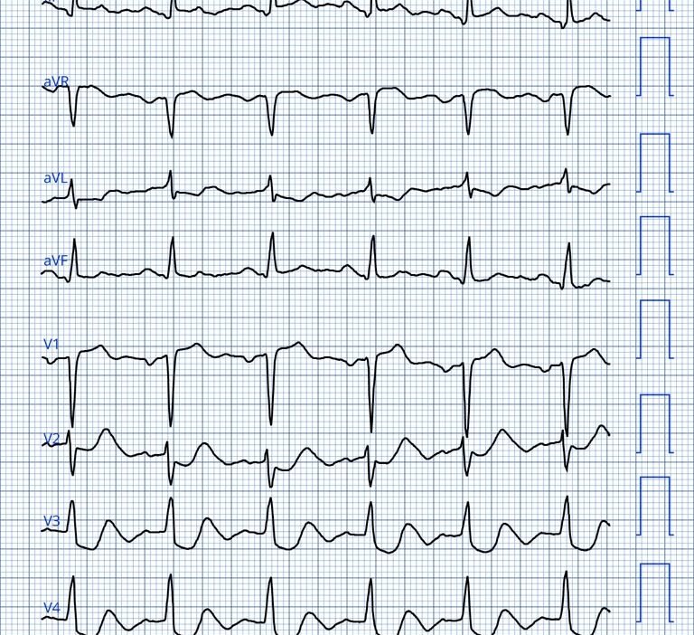
In Figure-1, to facilitate assessment — I’ve reproduced today’s initial ECG in an easier-to-assess format.
- The rhythm in ECG #1 is sinus with a normal PR interval, a narrow QRS — and a QTc that appears to be slightly prolonged. The frontal plane axis is normal — and there is no chamber enlargement.
Regarding Q–R–S–T Changes:
- Q waves of uncertain significance are seen in each of the inferior leds. Although these Q waves are not deep — they do appear to be slightly widened.
- R wave progression shows early transition (ie, The R wave becomes taller than the S wave is deep early, in between lead V2-to-V3).
The most remarkable changes relate to ST-T waves:
- ST elevation is noted in leads III, aVR and V1.
- As per the BLUE arrows — ST depression is noted in at least 7/12 leads! (in 8/12 leads if you count lead II). This ST depression is downsloping and marked in amount. This diffuse ST depression can not be missed!
= = =
Impression: As per Dr. Smith — Today’s initial ECG obtained from this 70-year old man with new persistent CP is diagnostic of acute OMI (= acute coronary occlusion).
- DSI (Diffuse Subendocarial Ischemia) is evidenced by the presence of ≥6-to-7 leads with ST depression in association with ST elevation in lead aVR.
- Acute inferior OMI is suggested by the Aslanger Pattern (ie, The presence of DSI in association with ST elevation in lead III).
- Acute posterior OMI is probably also present given the marked ST depression in leads V2,V3,V4.
- Given the ST elevation in lead V1 — right-sided leads would be indicated to rule out acute RV involvement.
- KEY Point: Even if treating clinicians fail to recognize these elements of DSI + ongoing infero-postero OMI (with possible RV involvement) — the marked diffuse ST depression can not be missed.
- P.S.: As per Dr. Smith — the diagnosis of “NSTEMI” is of no clinical utility, and is best avoided. But if clinicians refuse to give up this term — it is imperative that they at least be aware that: i) Many patients who are given the diagnosis of “NSTEMI” have cath-proven acute coronary occlusion in need of prompt cath and PCI; — and, ii) Regardless of what you put down in the chart as your admitting diagnosis — in a higher-risk patient who presents to the ED for new persistent CP — the ECG finding of diffuse marked ST depression mandates prompt cath to define the anatomy (and will almost certainly need prompt PCI).
= = =
Figure-1: I’ve labeled the initial ECG in today’s case.
= = =
= = =