Written by Dr. Ahmed Marai, who just graduated from Medical School in Iraq.
Edits by Smith and Grauer.
A 45-year-old man, an active smoker with no known past medical history, presented to a rural hospital with sudden-onset, severe retrosternal chest pain radiating to the left shoulder, which had started less than one hour prior to presentation. The pain was associated with diaphoresis and nausea. He described the chest pain as extremely severe.
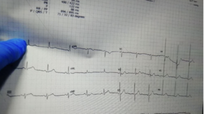
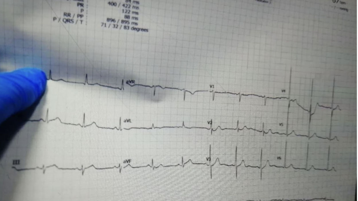
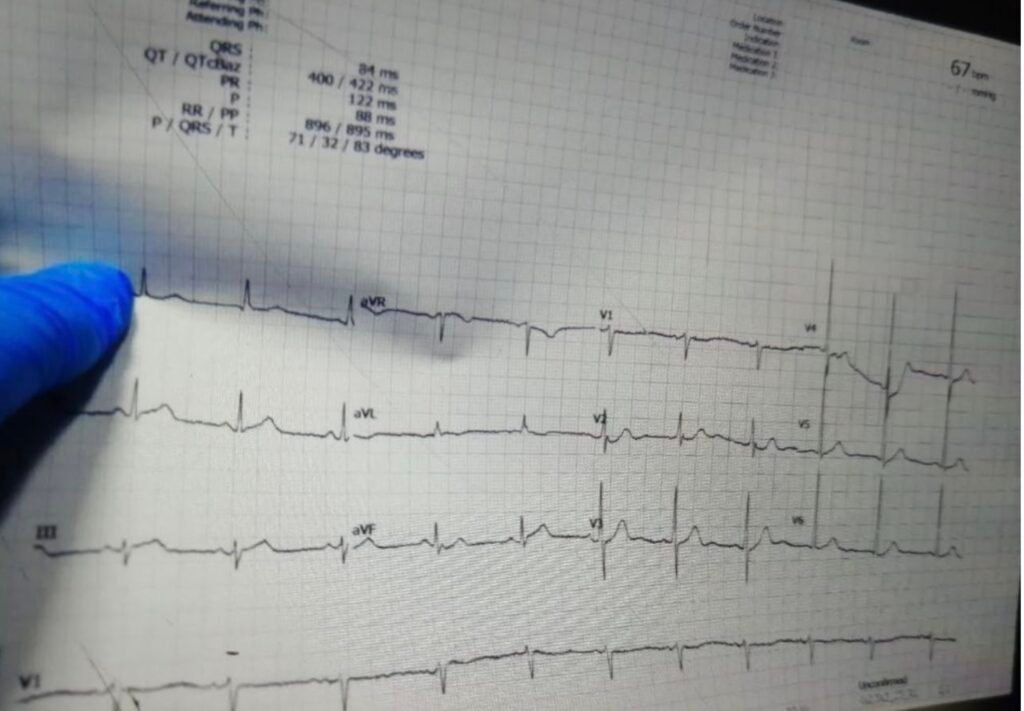
The patient’s vital signs were within normal limits. An initial 12-lead ECG was obtained in the emergency department. (Unfortunately this was the best quality tracing that could be obtained).
= = =
What do you think?
This ECG is diagnostic of inferior OMI. There is a hyperacute T-wave in lead III (it is not large by itself, but compared to the tiny QRS, it it very “large”, “bulky” and “inflated” and even has a high amplitude proportionally. There is reciprocal ST depression and T-wave inversion in aVL. ST depression in V2 and V3 strongly support OMI.
This ECG was interpreted by the treating physicians as a NORMAL ECG. Given the highly concerning clinical presentation, even without the ECG, acute coronary syndrome was suspected. The patient was managed with clopidogrel, subcutaneous low-molecular-weight heparin, and tramadol for analgesia.
His chest pain subsequently resolved (Smith: resolved due to tramadol? Do NOT use analgesics to manage ACS until you are committed to the cath lab!)
A second ECG was obtained, which was interpreted as “similar to the initial ECG”. (Unfortunately — Ahmed was unable to get a copy of this 2nd ECG.)
- NOTE: At presentation, the patient was experiencing severe chest pain. He was clutching his chest and describing what he was feeling as “crushing chest pain”. After recording the ECG — the treating physicians were shocked to see what appeared to be a normal ECG. Given the severity of the patient’s symptoms, they expected to see significant ST segment elevation. They wondered how a patient could have such severe chest pain with a normal ECG.
= = =
Smith: The ECG is not normal. I sent it to our EKG Nerdz group with no information, and everyone knew immedately that it is an infero-posterior OMI.
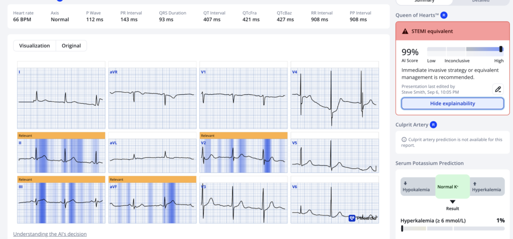
- Here is the PMCardio Queen of Hearts AI ECG Model interpretation:
= = =
So the cath lab should have been activated without even waiting for a troponin.
Serum cardiac troponin I was obtained and returned approximately one hour later.
- The hospital uses a qualitative troponin I assay — which reports the result as either positive or negative rather than providing a quantitative troponin concentration. The result returned negative.
- At that time, the patient was pain-free. The patient wanted to go home, as he thought he was okay. Despite urging by the treating physicians to remain for further evaluation — the patient refused, signed discharge paperwork, and left the hospital, believing that he was no longer at risk because the doctors had told him that his ECG was normal.
- Approximately one hour later, the patient returned to the emergency department in cardiac arrest. Resuscitation was unsuccessful, and he was pronounced dead.
- If you tell a patient that the ECG is normal, and you tell them their blood test is normal, they think they are fine and may resist any entreaty to stay. Giving pain medicines contributes to this false sense of security!
- See this post: What happens when you give morphine for chest pain in ACS? And what is pseudo-normalization of T-waves?
= = =
==================================
MY Comment, by KEN GRAUER, MD (9/9/2026):
As discussed above — Today’s patient is a 45 yo man who presents with a 1-hour history of severe “crushing” retrosternal CP (Chest Pain), in association with nausea and diaphoresis.
- As evident from the above discussion of today’s case — providers realized that this simple history (of new severe “crushing” CP) immediately placed this patient in a high-risk group for having an acute cardiac event.
- What providers did not fully appreciate — was that because today’s patient falls in a higher-risk group — we need to lower our threshold for determining what constitutes an “abnormal” ECG.
- NOTE: As emphasized many times in Dr. Smith’s ECG Blog — an initial high-sensitivity Troponin may be negative despite the patient having an acute occlusion MI.
- An initially negative high-sensitivity Troponin value in patients with acute coronary occlusion occurs more commonly than many providers appreciate!
= = =
Today’s initial ECG: Technical Challenges
The clinical reality is that the technical quality of an acute ECG is not always optimal. There are many reasons for this, given that an acute patient may be having difficulty breathing, or may be in severe pain, restless, agitated, shivering, tremulous, or otherwise unable to remain still.
- As per the well-known IT truism: “Poor data in = poor data out”. That said, the clinical reality is that for a variety of reasons — optimal quality ECG tracings are not always feasible. Today’s case provides an insightful example of how to make the best of what we have available!
Among the technical challenges in today’s tracing are the following:
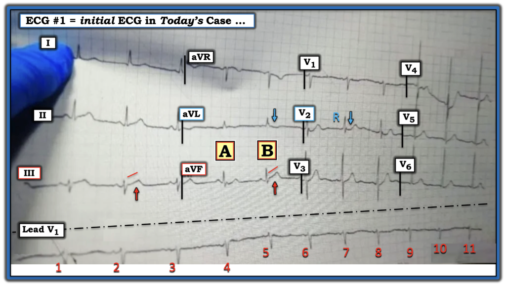
- i) The tracing is markedly slanted (with downward inclination of the 12-lead — and upward inclination of the long lead rhythm strip, as shown in Figure-1). This results in significant distortion of measurements — as well as distorting what to use for the ST segment baseline from which we determine ST elevation or depression (We essentially never see a series of beats in a row with a constant horizontal baseline).
- ii) In addition to the marked inclination of the tracing in Figure-1 — there is artifact operative in the 2 QRS complexes seen in the simultaneously-recorded augmented limb leads ( = beats #4 and 5 — which I’ve labeled “A” and “B” ). This raises the question as to which beat (A or B?) represents the true ST-T wave in lead aVF?
= = =
My Thoughts on Figure-1:
In Figure-1 — Note the baseline wander in the long lead V1 rhythm strip at the bottom of the tracing. Distortion of this tracing is so marked — that I suspect the overall rhythm is fairly regular despite what seems like marked discrepancy in the R-R intervals of the 11 beats on this tracing.
- But — Note that the baseline in the long lead rhythm strip does become almost horizontal beginning with beat #5. This supports my suspicion that beat #5 ( = complex “B” in Figure-1) represents the true appearance of the ST-T wave in lead aVF.
- This supposition makes sense — because the ST segment straightening with subtle-but-real ST elevation that we see for “B” in lead aVF looks identical to the ST-T wave appearance for beat #2 in lead III.
- And — complex “B” ( = beat #5) in lead aVL suggests there is the perfect picture of reciprocal ST depression (the BLUE arrow in lead aVL) — that in this patient with “crushing” new CP all but confirms an acute inferior OMI!
- As I always do when contemplating the possibility (in this case, probability) of acute inferior OMI — I look for potential QRST changes of posterior OMI (since the common blood supply to inferior and posterior LV walls in most patients so often results in both inferior and posterior OMIs occurring together).
- ECG indication of acute posterior OMI is forthcoming in lead V2 from: i) Loss of the slight, upward sloping ST elevation that we normally see in lead V2 (BLUE arrow in this lead); — and, ii) The presence of a predominant R wave already in lead V2 (this predominant R wave being a Q wave “equivalent” with posterior OMI).
= = =
Figure-1: I’ve reproduced and labeled today’s ECG.
= = =
Final Concepts:
Providers in today’s case had difficulty accepting how a patient with such severe new CP could have such a “normal-looking” ECG.
- As per Dr. Marai — Today’s ECG is not normal. Instead, in this patient with new severe CP — it is diagnostic of acute infero-postero OMI until proven otherwise (and should mandate prompt cath for PCI ).
- The reasons that an ECG may sometimes look relatively normal despite ongoing acute coronary occlusion are: i) Simply “because” (ie, Because sometimes this happens); — ii) Because “beauty is in the eye of the beholder” (What appeared “normal” to providers in this case is not “normal” in the eye of an expert interpreter); — and, iii) Because as part of the normal evolution of acute coronary occlusion — there is often a pseudo-normalization period in which the ECG may look relatively “normal” (ie, if an ECG is recorded in between the period of initial ST elevation and the period of reperfusion when ST-T waves are depressed and T waves are inverted).
PEARL #1: An all-too-often-ignored clinical clue that your patient may be in the “pseudo-normalization” period — is if at the time a given ECG is recorded, the patient’s CP has decreased or resolved.
- But if you do not routinely ask the patient about the presence and relative severity of their symptoms each time you record an ECG — then you will never profit from the invaluable information forthcoming when you correlate each ECG recorded with the presence and severity of the patient’s CP.
- And after you ask the patient about their symptoms — Write this down on the actual ECG (ie, that the patient’s CP was still 9/10 or 1/10 or whatever at the time that ECG was recorded). Because, if you do not write the patient’s pain score on the ECG — the clinical reality is that you will never know later if the patient’s CP was increasing, decreasing or the same at the time that ECG was recorded.
PEARL #2: Although this next PEARL should seem obvious — it is all-too-commonly ignored. If for whatever reason there are “technical challenges” to the interpretation of a given ECG in a patient with suspected OMI (be this from baseline wander, artifact, patient movement, or other cause) — Simply repeat the ECG immediately!
- I am continually disappointed by how often this is not done. Much of the time when you alert the technician to try again by immediately repeating the ECG — You’ll be able to improve the technical quality enough to facilitate a more reliable interpretation (and you’ll convey to the technician the need to automatically try again when there are technical challenges to always obtain the “best possible” recording for your interpretation).
= = =
= = =