Written by Matthew McArthur
A 60 year old patient was brought in to the Emergency Department at 3am by EMS with one hour of crushing chest pain and diaphoresis. EMS ECG was “STEMI negative” so the patient was brought to a hospital without PCI capability.
Unfortunately, we do not have access to that ECG.
Patient received aspirin, and multiple doses of nitro and morphine with EMS with ongoing severe 8/10 pain.
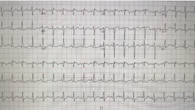
Here is the initial ECG in the ED.
The ECG machine interpretation was:
- Sinus Rhythm
- Right Bundle Branch Block
- Borderline ST elevation, lateral leads
What do you think?

.
.
.
Looking at the ECG: There is a Right Bundle Branch Block. There is contiguous, convex ST elevations ballooning into wide, broad-based T waves in the lateral precordial leads (V4-V6) with reciprocal ST depression in V1-V2. This is diagnostic for lateral wall ischemia.
This STE does not meet STEMI millimeter criteria (there is 1 out of 4 complexes in which it does, due to baseline wander). STE criteria measure STE at the J-point relative to the PQ junction (QRS onset).
In V1, there is ST depression. Some ST depression is expected in RBBB, discordant to the wide tall R’-wave. The R’-wave amplitude here is quite small, though, so one might not expect much ST depression.
Reverse precordial swirl
This is the reverse of the Precordial Swirl Pattern which is well-described in previous blog posts. Ischemia in the septum (V1-V2) can cause reciprocal ST depression in the lateral leads (V5-V6) (precordial swirl). Here we have lateral wall ischemia (V4-V6) causing reciprocal ST depression at the septum (V1-V2).

A 15 lead ECG was also completed which did not show any ST elevation in the posterior leads.
Queen of Hearts had no doubt about the diagnosis of Occlusion MI, or the culprit artery:

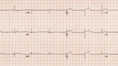
A previous ECG from a few years ago was available for comparison – confirming the lateral STE/HATW and reciprocal changes were new:

I contacted the on-call STEMI Physician. Patient was accepted for emergent transfer for primary PCI. Cath lab revealed a culprit 99% proximal occlusion of the obtuse marginal artery. PCI went well.
Take away
This case illustrates the risk of over-reliance on STEMI millimeter criteria and ECG machine interpretation, and has a few key important factors to review:
- We do not have that first ECG. We do not know if it showed OMI in spite of not showing STEMI. However, a 60 year old with crushing chest pain should be brought to the nearest PCI capable facility no matter the ECG findings.
- STE in the context of RBBB – can be overcalled or undercalled due to the secondary repolarization changes seen in right bundle
- contiguous, concordant, <1mm STE in the lateral precordial leads
- convex morphology of STE, ballooning into wide, broad-based T waves
- reciprocal STD, reverse precordial swirl pattern
- usefulness of reviewing prior ECG when available
The ECG paints a picture based on the confluence of all these factors. Understanding the nuances of ECG interpretation will allow greater confidence in diagnosing acute MI that does not meet STEMI millimeter criteria, and in differentiating acute MI from STEMI mimics.
Queen of Hearts AI has been trained on millions of ECGs, and uses pattern recognition to detect OMI, including OMIs like this one that may be missed by conventional STEMI criteria or ECG machine interpretation. If EMS was using QOH, this patient would likely have been transported immediately to the PCI centre resulting in significantly lower door to balloon time and timelier reperfusion for better longterm outcomes.
= = =
==================================
MY Comment, by KEN GRAUER, MD (9/11/2026):
Today’s case by Dr. McArthur provides yet one more illustration of why the STEMI protocol is simply not good enough. Despite new-onset severe CP (Chest Pain) requiring multiple doses of Nitroglycerin and Morphine — this patient was brought by EMS to a hospital without PCI capability because, “his initial ECG was STEMI negative”. Among the reasons for the fallacy of this approach are the following:
- A history of new-onset CP that is sufficiently severe to awaken the patient from sleep, and persistent enough such that multiple doses of Nitroglycerin and Morphine administered by EMS providers failed to relieve his CP — immediately places this patient at a higher-risk likelihood for needing prompt cath with PCI regardless of what the initial ECG shows.
- Even in patients who do eventually satisfy STEMI criteria on ECG — these criteria are often not yet present on the initial ECG (sometimes only developing many hours later).
- As we’ve repeatedly shown on Dr. Smith’s ECG Blog (and as we show again in today’s case) — acute coronary occlusion (ie, an OMI) may be readily detected on the initial ECG despite a lack of definitive STEMI-positive criteria.
= = =
Today’s initial ECG:
I suspect that the reason today’s initial ECG was not recognized as indicative of an acute OMI — is the atypical nature of this 1st tracing.
- For clarity and ease of comparison in Figure-1 — I have reproduced and labeled this initial tracing, and placed it side-by-side with a previous ECG from this patient.
= = =
Atypical features of today’s initial ECG include the following:
- The QRS complex in lead V1 in the initial tracing is not only small — but it lacks the familiar triphasic rsR’ (in whch there is s wave descent below the baseline — and a clearly defined “taller right rabbit ear”). Instead — the s wave in lead V1 barely reaches the baseline, and the R’ wave is rounded and diminutive, with no sign of anything resembling a triphasic complex in other anterior leads.
- The QRS complex in lead I manifests a qrS configuration that lacks the R wave amplitude that is typically seen with RBBB in the absence of an associated posterior hemiblock.
- The terminal s wave in lead V6 does not look wide.
- The downsloping ST-T wave depression in lead V1 that is characteristic of typical RBBB — is abbreviated by a barely depressed, rounded ST segment that disappears by lead V2.
- In short — I interpreted this initial ECG as showing “RBBB” (Right Bundle Branch Block) not because QRS morphology “looks like” a typical RBBB — but rather because of a lack of any better description for the atypical morphology with slight QRS widening that is seen in this tracing.
= = =
Figure-1: Comparison between the 2 ECGs in today’s case.

= = =
Features Indicative of an Acute OMI:
Despite the atypical features of RBBB — in this 60-year old man with new-onset severe CP — the initial ECG is diagnostic of an acute OMI until proven otherwise because:
- There clearly is coved ST elevation in leads V4,V5,V6 — thereby indicative of a “lateral” OMI (RED arrows in Figure-1). Although hard to delineate the J-point because of the coved (“frowny” shape) ST segments in these 3 consecutive lateral chest leads — I thought the J-point was a millimeter elevated.
- The ST-T wave in lead V2 is flattened. Although marred by baseline artifact and the somewhat variable appearance of the 3 complexes seen in lead V2 — the 3rd complex in this lead does appear to show downsloping ST depression (BLUE arrow in lead V2) — which rather than a pure “reciprocal” change, I attributed to “posterior” OMI.
Impression: In this 60-year old man with new-onset severe CP — I interpreted the initial ECG in today’s case as showing acute postero-lateral OMI, consistent with a “culprit” artery in the OM (Obtuse Marginal) branch of the LCx (Left Circumflex) coronary artery.
- P.S.: Although ST-T waves in the limb leads of ECG #1 are non-diagnostic — larger-than-expected Q waves (considering tiny size of the R waves that follow) are present in leads I and aVL (YELLOW arrows). Especially given what appears to be slight widening of the Q waves in lead aVL — I initially wondered if these Q waves might represent acute high-lateral lead changes.
The above said — on retrieving a previous ECG from today’s patient, a more complete picture immediately became evident:
- Similar appearing high-lateral Q waves (that are subtly widened in lead aVL) are seen on this previous ECG — such that these Q waves are not a new finding.
- A similar RBBB (or incomplete RBBB?) pattern was present in the earlier tracing.
- On looking at ECG #2 (the previous tracing) — there is no doubt that the lateral chest lead ST elevation in leads V4,V5,V6 of today’s initial ECG is new!
- The ST segment flattening I perceived in lead V2 of today’s initial ECG is not present in the earlier tracing (Instead — there is subtle slight upsloping of the ST segment in ECG #2).
Bottom Line: Any doubt that might have been present about the acuity of today’s initial ECG — should have been immediately discounted as soon as the previous ECG was retrieved for comparison!
= = =
= = =