A middle-aged man called EMS for chest pain.
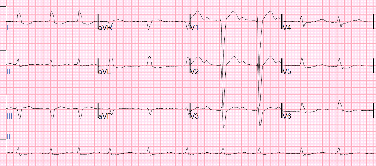
This prehospital ECG was recorded:
 |
| Obvious Anterior STEMI due to proximal LAD occlusion (with STE in aVL and reciprocal STD in inferior leads). |
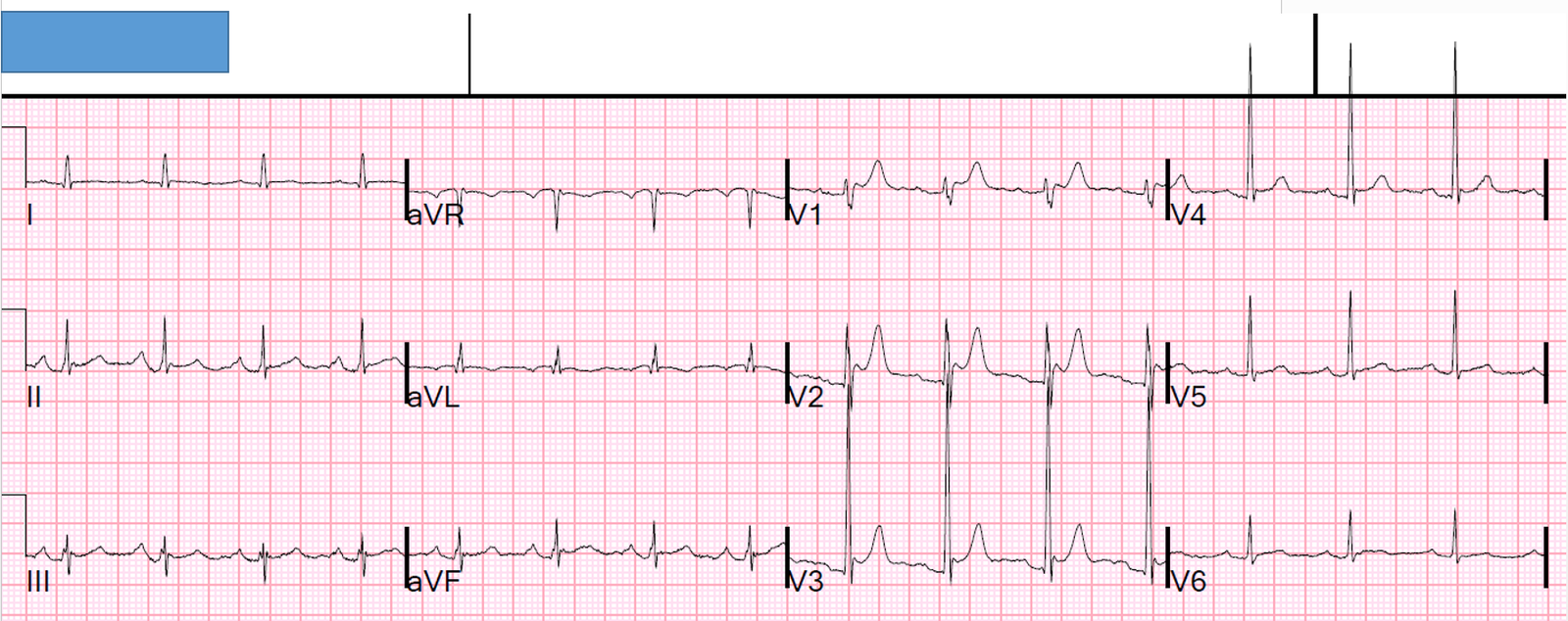
On arrival to the ED, this ECG was recorded 10 minutes later:
 |
| Almost all STE is gone, but the hyperacute T-waves remain |
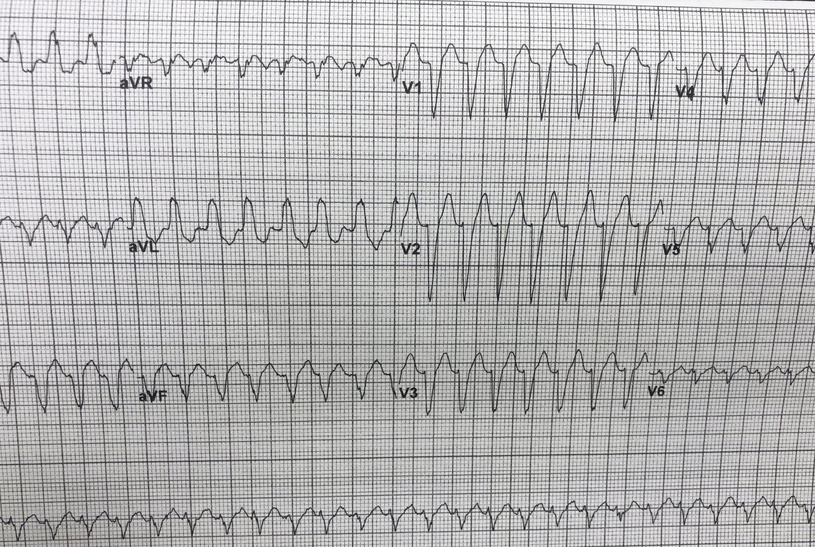
While waiting for the cath team, this was recorded 30 minutes after 2nd ECG:
 |
| Now there are classic de Winter’s T-waves. |
This shows the dynamic nature of coronary thrombus. Presumably, the thrombus had autolysis to a very small degree, allowing a trickle of blood flow through the LAD, enough to eliminate the ST elevation.
The patient was found to have a 100% proximal LAD occlusion.
By the time of the angiogram, which is never at the same the time as the ECG, there was not even a trickle of blood.
See this related post, with discussion of de Winter’s T-waves: