Written by Pendell Meyers with edits by Smith.
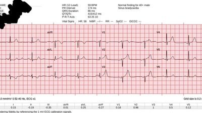
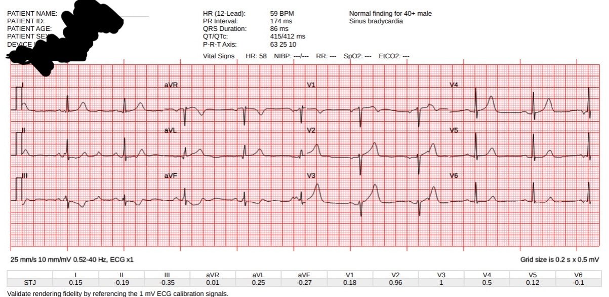
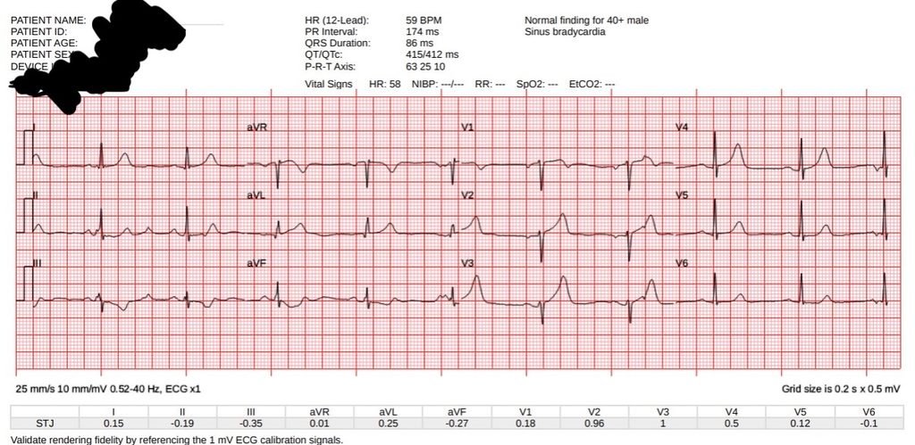
A man in his 40s called EMS for acute onset chest pain. EMS arrived and recorded vital signs within normal limits, and then this ECG during active chest pain:
What do you think?
The EMS machine algorithm lead mentions the sinus bradycardia at 59 bpm, but otherwise calls this ECG “normal.”
I disagree strongly! I think it is ~100% diagnostic of acute total LAD occlusion. Ischemic STE in V1-V3 (less than STEMI criteria), poor R waves there, and HATW in V2, V3, V4, I, aVL. Precordial swirl pattern is completed by the STD in V6. Reciprocal STD and depressed T wave in III and aVF.
Smith: Another very specific sign which I always look for when wondering about a hyperacute T-wave in V2 is down-up T-wave in leads III or aVF. Sure enough, there is a down-up T-wave in aVF.
PMcardio Queen of Hearts AI ECG Model interpretation (now FDA approved):
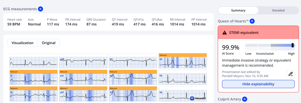
The Queen agrees.
Code STEMI was activated, despite no STEMI criteria (gasp!)
The cardiologist was “not impressed” with the ECG, but did not cancel the cath lab.
A repeat ECG was performed:
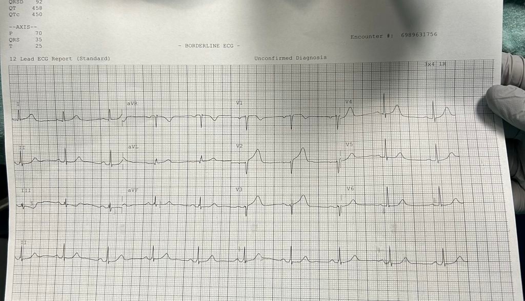
Slightly easier, but still not STEMI criteria.
The ED physician recorded this echocardiogram (parasternal short axis) which was interpreted as an anterior wall motion abnormality:
If the team had waited for the first troponin, it would have been within normal limits at 7 ng/L (high sensitivity troponin I, URL <20 for men).
But they did not wait, they went quickly to the cath lab:
The operating Cardiologist documented a total LAD occlusive culprit lesion and performed PCI with “excellent angiographic result”.
The door to balloon time was under 60 minutes!
The next two troponins were both greater than 25,000 ng/L.
Formal echo confirmed the wall motion abnormality.
The patient did well during the visit and was discharged.
What should the discharge diagnosis be?
No STEMI criteria ever on any ECG. Yet “treated as STEMI.” So, final diagnosis: “STEMI.”
Smith: We showed that the final diagnosis of “STEMI” vs. “NSTEMI” has almost nothing to do with presence of STEMI millimeter criteria, or even with presence of occlusion. Interventionalist diagnosis of STEMI vs. NSTEMI is most closely correlated with door-to-balloon time.
= = =
Learning Points:
Do not trust conventional ECG machine algorithm interpretations. Become an expert yourself, or use a validated and approved AI model like the Queen of Hearts, or both!
The disease of acute coronary occlusion is not well defined by ST millimeters on the ECG. Please, be a real doctor and try to find the disease even when its not “barn door” easy on the ECG.
Hyperacute T waves are an important piece of how OMI can be found faster than standard care in 2026.
Smith: I have 65 cases of OMI on this blog for which the conventional algorithm says the ECG is totally normal, yet I could see that it was OMI (and it was indeed OMI). We tested Queen of Hearts on this group. She was 81% sensitive for OMI.
- Karim S…..Smith SW. Artificial Intelligence Detection of Occlusive Myocardial Infarction from Electrocardiograms Interpreted as “Normal” by Conventional Algorithms
- Meyers HP et al…..Smith SW. Hyperacute T Waves Are Specific for Occlusion Myocardial Infarction, Even Without Diagnostic ST-Segment Elevation (full text)
See how HATW identify LAD Occlusion (as does Smith and Queen):
- Meyers HP…..Smith SW. Failure of standard contemporary ST-elevation myocardial infarction electrocardiogram criteria to reliably identify acute occlusion of the left anterior descending coronary artery. (also full text)
= = =
======================================
MY Comment, by KEN GRAUER, MD (11/11/2026 ):
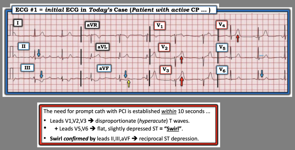
Today’s case should be an EASY one — as it literally took me less than 10 seconds on learning that this patient has acute onset CP (Chest Pain) — to appreciate that today’s initial ECG shows:
- Hyperacute T waves in leads V1,V2,V3 + ST segment flattening with slight depression in leads V5,V6 (Should take less than 5 seconds to see this … ).
- And, there is reciprocal ST depression in the inferior leads (Should take no more than another 5 seconds to see this … ).
= = =
Breaking these less than 10 seconds down …
We’ve reviewed numerous cases of Precordial “Swirl“ on Dr. Smith’s ECG Blog (See My Comment at the bottom of the page in the October 15, 2022 post — for review of the KEY features for prompt recognition of Swirl). There is no magic to making the above diagnosis. It is simply “fact until proven otherwise”, as shown in countless cases with cath-documented proof of it validity.
- There is simply no way that the T waves in leads V2,V3,V4 in Figure-1 are “normal” in a patient who presents with acute CP. Instead — our “eye” should be immediately captured by the clearly disproportionately increased T wave in lead V3, that can easily “swallow” within it the diminutive QRS in this lead.
- In the context of this disproportionately enlarged T wave in lead V3 — the T waves in neighboring leads V2 and V4 are also disproportionately enlarged ( = overly “bulky” ==> hyperacute).
- The diagnosis of “Swirl” until proven otherwise is then made by the ST segment flattening with slight depression in lateral chest leads V5,V6.
- Seeing clear evidence of reciprocal ST depression in each of the inferior leads ( = leads II,III,aVF) immediately confirms our conclusion of the need for PCI until proven otherwise by prompt cath.
= = =
The finer details in Today’s CASE:
There are finer details in virtually all leads on today’s initial ECG — but as regular readers of Dr. Smith’s ECG Blog will attest to — confidence in the diagnosis of Swirl until proven otherwise should be arrived at within seconds in this patient with acute CP simply from the above findings. For teaching purposes — I’ll add:
- There is poor R wave progression ( = tiny initial r waves in leads V1,V2,V3 — which if this is a new finding — suggests loss of anterior forces from ongoing anteroseptal infarction).
- As is typically the case with Swirl — the ST-T wave appearance in lead V1 is not normal (In Figure-1 — the ST segment in this lead is straightened, elevated — and with terminal negativity of the up-down T wave in this lead). Knowing that leads V2,V3,V4 are hyperacute facilitates my knowing that the ST-T wave in lead V1 is also abnormal.
- Lead V5 makes for an interesting “transition” lead — as it shows vestiges of T wave hyperacuity (a T wave bulkier and “fatter”-at-its-peak than it should be) as the transition is made from anterior lead hyperacute T waves to the reciprocal ST flattening and depression characteristic of Swirl that we see in lead V6.
- As emphasized by Dr. Smith — the tell-tale down-up T wave in lead aVF (YELLOW arrow) — confirms hyperacuity of the anterior T waves. Although marred by artifact — another down-up T wave is probably also present in lead III.
- Inferior lead reciprocal ST depression is further supported in lead aVL (this T wave in aVL being overly “bulky” — and a near mirror-image opposite picture of the deep inverted T wave in lead III).
- The other high-lateral limb lead = lead I — also shows a bulkier-than-expected hyperacute T wave.
Bottom Line: The cardiologist in today’s case was “not impressed” by today’s initial ECG — but at least he/she did not cancel the cath lab activation. Hopefully that cardiologist will in the future realize that the need for prompt cath in cases like this is readily apparent within less than 10 seconds of knowing what to look for.
= = =
For clarity in Figure-1 — I’ve labeled today’s initial ECG.
= = =
= = =