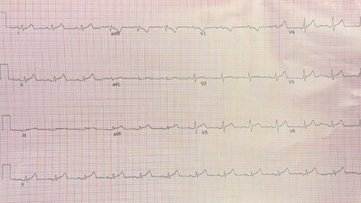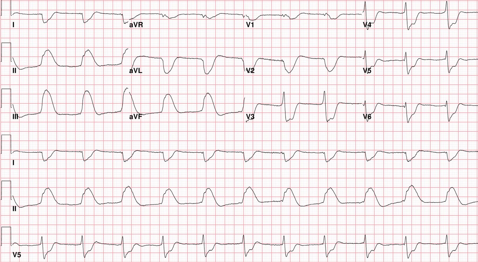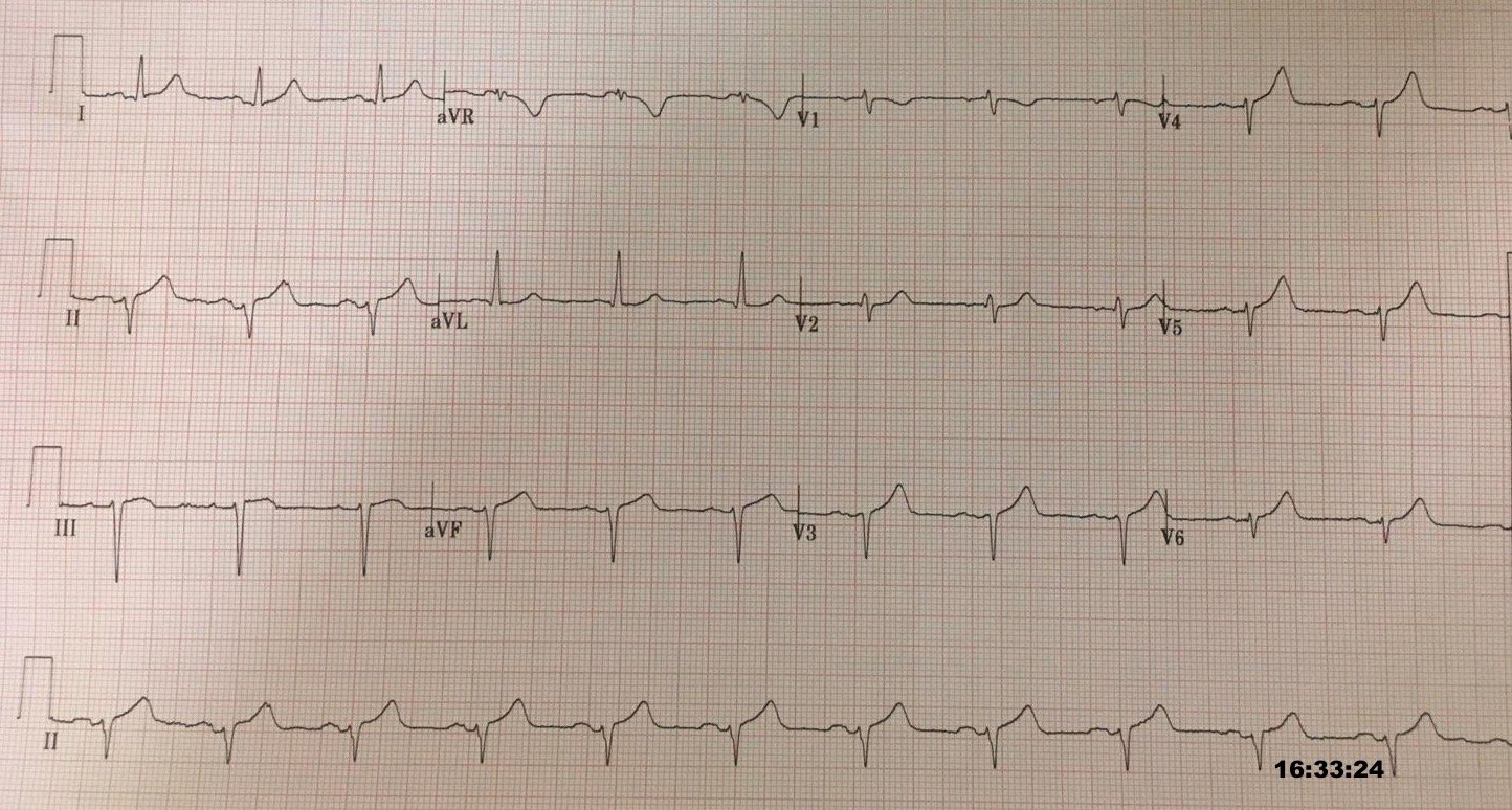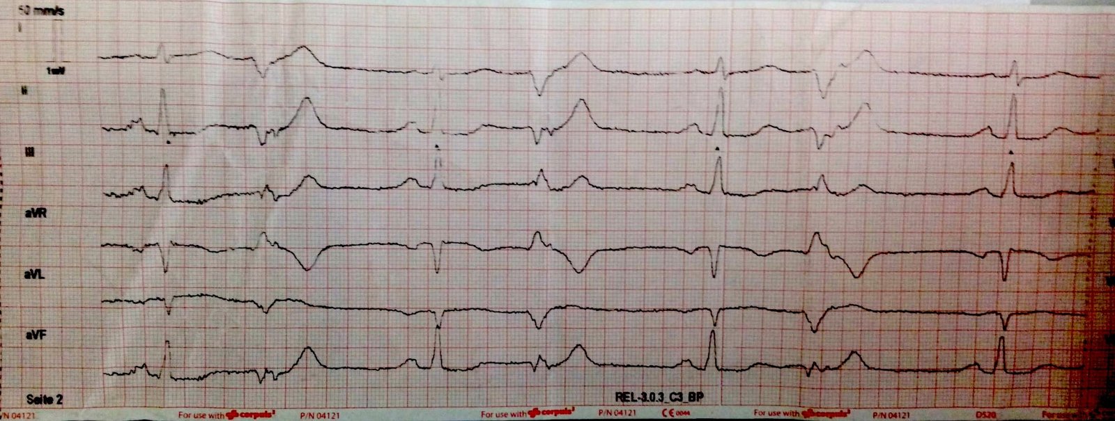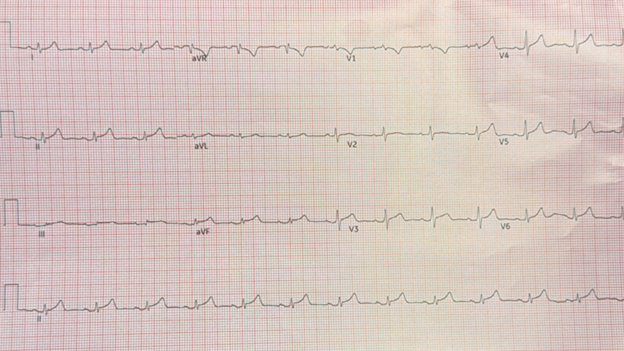
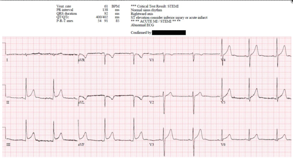
A former resident was working in triage when this ECG was handed to her, recorded on a 73 year old chest pain patient.
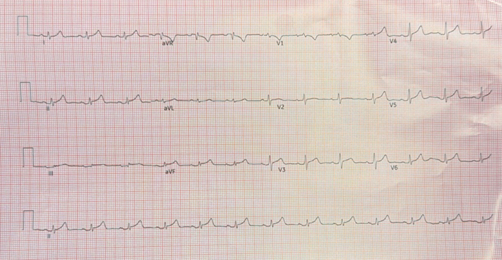
— What do you think? —
= = =
= = =
The conventional algorithm output was *** Acute MI/STEMI ***.
This former resident saw the ST Elevation, but was skeptical of OMI because “The T-wave morphology just did not look right for OMI.”
Kudos to her!
I would add that the upright T-wave in aVL makes the inferior ST depression much less worrisome, even with that slight ST depression in aVL which can be seen in early repol (but is very rare in pericarditis). Nevertheless, sometimes early repolarization can manifest with both STD and reciprocally inverted T-wave in aVL. I will post those images at the bottom.
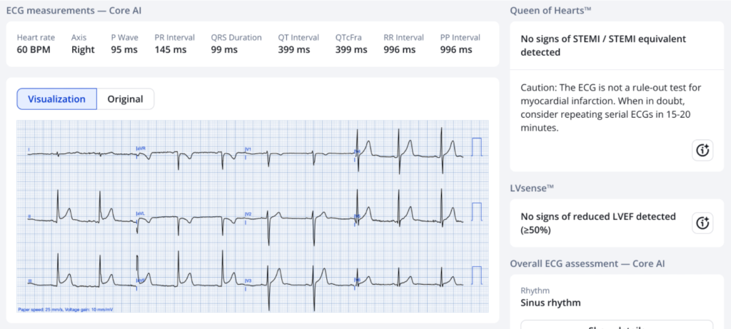
She confirmed her suspicions using the PMCardio Queen of Hearts AI ECG Model:
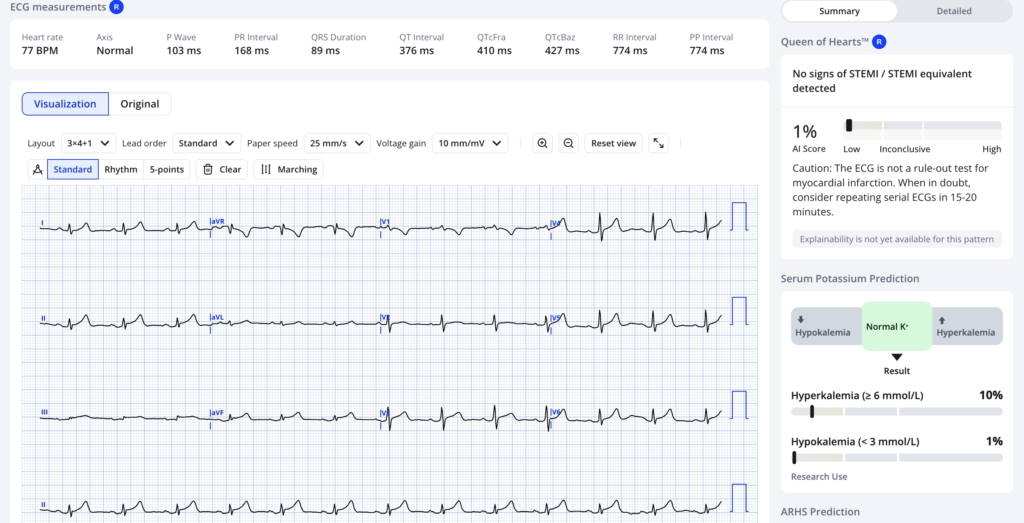
= = =
Our Model that is not yet available gives OMI Mimic probabilities. Here they are:
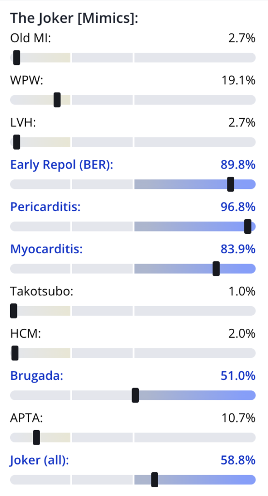
= = =
A 73 year old with chest pain has a relatively high pretest probability of OMI, and a lower likelihood of early repolarization, so one should be very careful about dismissing this ECG.
She did not activate the cath lab, but appropriately brought the patient to the ED, to another MD, for further investigation.
2 troponins were 7 ng/L.
It is uncertain if more serial ECGs were done, but this should be done every 15-20 minutes for persistent chest pain, as one must be CERTAIN that such an ECG does not represent OMI.
D dimer was slightly elevated, but CT pulmonary angiogram was negative.
Can the PMcardio Queen of Hearts AI ECG Model decrease false positive activations?
In this study from JACC: Cardiovascular Interventions among 1050 cath lab activations, the Queen decreased false positive activations from 42% using standard of care (which includes HPI, medical history, exam, and ECG) to 8% (Queen using ECG only), while INCREASING sensitivity on the first ECG from 71% to 92%
Sensitivity AND specificity were far better: 92% vs. 71% for sensitivity. 81% vs. 29% for specificity.
In another study of prehospital cath lab activations, the Queen decreased false positive activations from 69/117 to 29/117, a 58% decrease, while not missing any of 48 true OMI.
In yet another study of 242 patients with myocarditis or pericarditis, the Queen correctly classified 230 (95.0%) as Not OMI, far better than ST Elevation millimeter criteria (78.1% specificity) and better than all large language models (specficity 27%-70%)
= = =
Epilog
Final diagnosis was the most common chest pain diagnosis: unknown etiology. It is possible that it is pericarditis, but benign chest pain and early repol are by far most likely.
= = =
Another instructive case
See this case of early repol with BOTH STD in aVL and inverted T-wave. Dr. Smith and Queen both (blinded) recognized it as Not OMI:
Queen:
= = =
======================================
MY Comment, by KEN GRAUER, MD (10/27/2026 ):
It’s insightful to occasionally see suspicious ECGs turn out not to be the result of an acute OMI:
- Confession: I was not comfortable concluding solely from today’s initial ECG that this tracing was benign. Instead — I was comfortable with the wisdom and clinical reality of knowing that sometimes, more than a single ECG is needed for optimal clinical decision making.
- The “good news“ — It’s OK not to be certain from a single ECG if a given patient is or is not in process of evolving an acute OMI. That’s because i) The history can guide us (depending on how worrisome the history sounds for an acute cardiac etiology); — ii) Finding a prior ECG on the patient may provide insight as to whether potential concerning ECG findings are new or old; — iii) Serial ECGs looking for “dynamic” ECG changes often provide the answer as to whether or not suspicious ECG findings are evolving, as they usually do with acute OMI (and with ongoing chest pain — “dynamic” ST-T wave changes may be seen in as little as 10-15 minutes!); — iv) Two or more negative hs-Troponins is reassuring (as was seen in today’s case); — and, v) Stat bedside Echo can be reassuring if no localized wall motion abnormality is seen in association with ongoing symptoms.
- Bottom Line: It’s OK not to be certain from the intial ECG if a given patient is or is not in process of evolving an acute OMI — because we have tools that can help us within a short period of time to determine the likelihood of acute OMI.
= = =
Why I Found Today’s Case to be Difficult …
Several features made today’s case difficult for me:
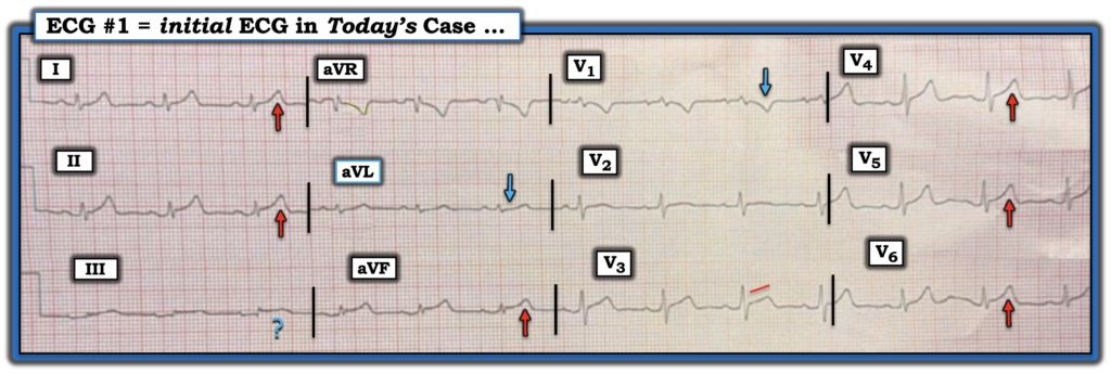
- I found the initial ECG deceptive — if for no other reason than how tiny QRS voltage is in so many leads. In my effort to optimize visualization — I reduced spacing between the leads and I was able to slightly increase size of this tracing (See my Figure-1 below).
- ECG findings do not localize in today’s tracing the way they usually do with acute OMI. The ST-T wave in lead II looks more like the ST-T wave in lead I than in lead III (a feature more characteristic of pericarditis than of inferior OMI). Nevertheless, despite minuscule size of the QRS complex in lead III — Doesn’t the BLUE ? in lead III look suspicious for an acute ST elevation?
- Lead aVL is puzzling. Once again, tiny size of the QRS in this lead is deceiving. I was concerned by subtle-but-real ST depression (BLUE arrow in this lead) — and as I strained my eyes looking multiple times at lead aVL, I imagined the T wave to be disproportionately enlarged compared to the less than 2 mm R wave in this lead.
- In the chest leads — I thought the ST coving and size of the inverted T wave in lead V1 was unusual — the ST segment in lead V3 looked straightened, as well as being elevated — and the T waves in leads V4,V5,V6 looked taller than expected given modest size of the R waves in these leads.
- Bottom Line: I found today’s ECG to be difficult to interpret in this 73-year old patient who presented to the ED for chest pain. As I contemplated the case in the comfort of my dual large-screen monitors and stressless office chair — I was not comfortable ruling on OMI vs non-OMI on the basis of this single ECG. I felt that if I was there at the bedside — I’d need more information before ruling on this initial tracing.
= = =
Figure-1: I’ve reproduced today’s initial ECG, adjusting some features to optimize visualization.
= = =
Final Thought …
Dr. Smith concludes discussion of today’s case with reference to the May 23, 2022 post, in which Dr. McLaren asks us to decide without clinical context — Which one or more of 4 ECGs is diagnostic of acute OMI?
- I just now once again read through the above cited excellent post by Dr. McLaren that I had commented on 4 years ago, forcing myself (without looking at what I had said in 2022) to commit to an “acute OMI or no acute OMI” diagnosis based solely on the initial ECG without clinical context. Doing so convinced me of the wisdom and clinical reality that sometimes more information is needed before we can be comfortable rendering a diagnosis.
- P.S.: For any readers with a moment of time who might want an additional challenge — the above cited May 23, 2022 post is timeless in its wisdom and insight.
= = =
= = =