This case was sent by a former resident.
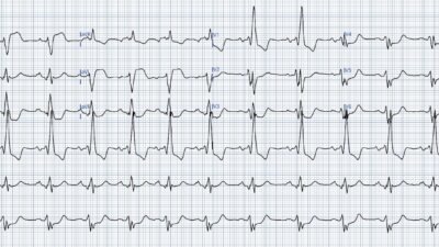
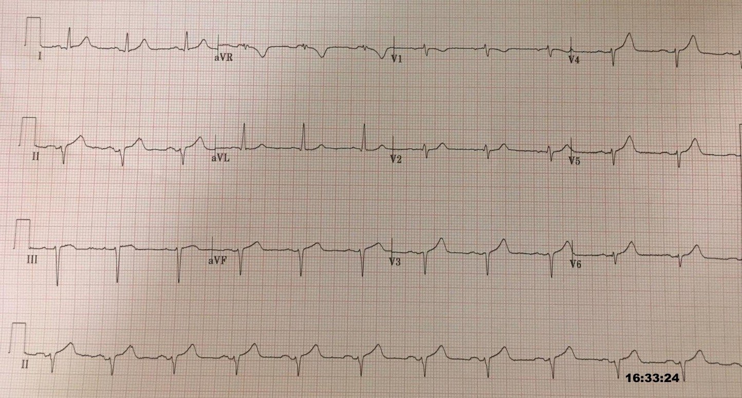
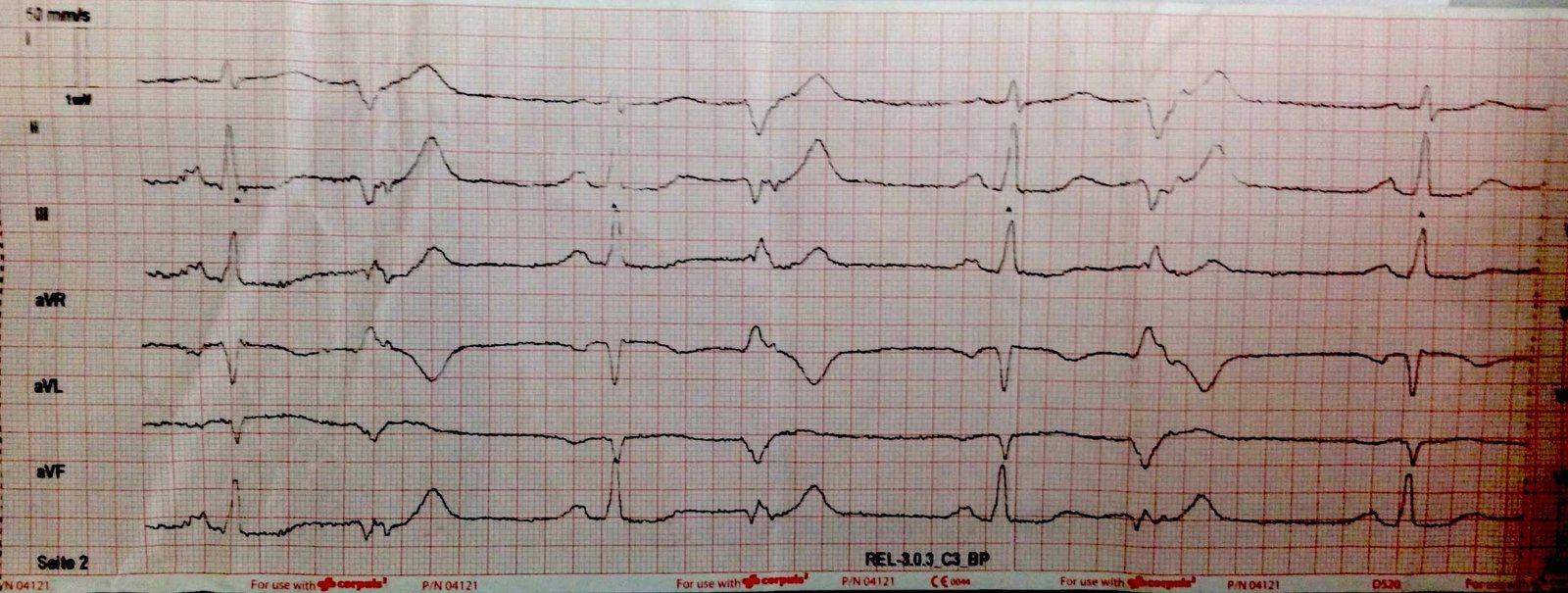
This middle aged woman with history of coronary disease and CABG presented with Chest pain:
What do you think?
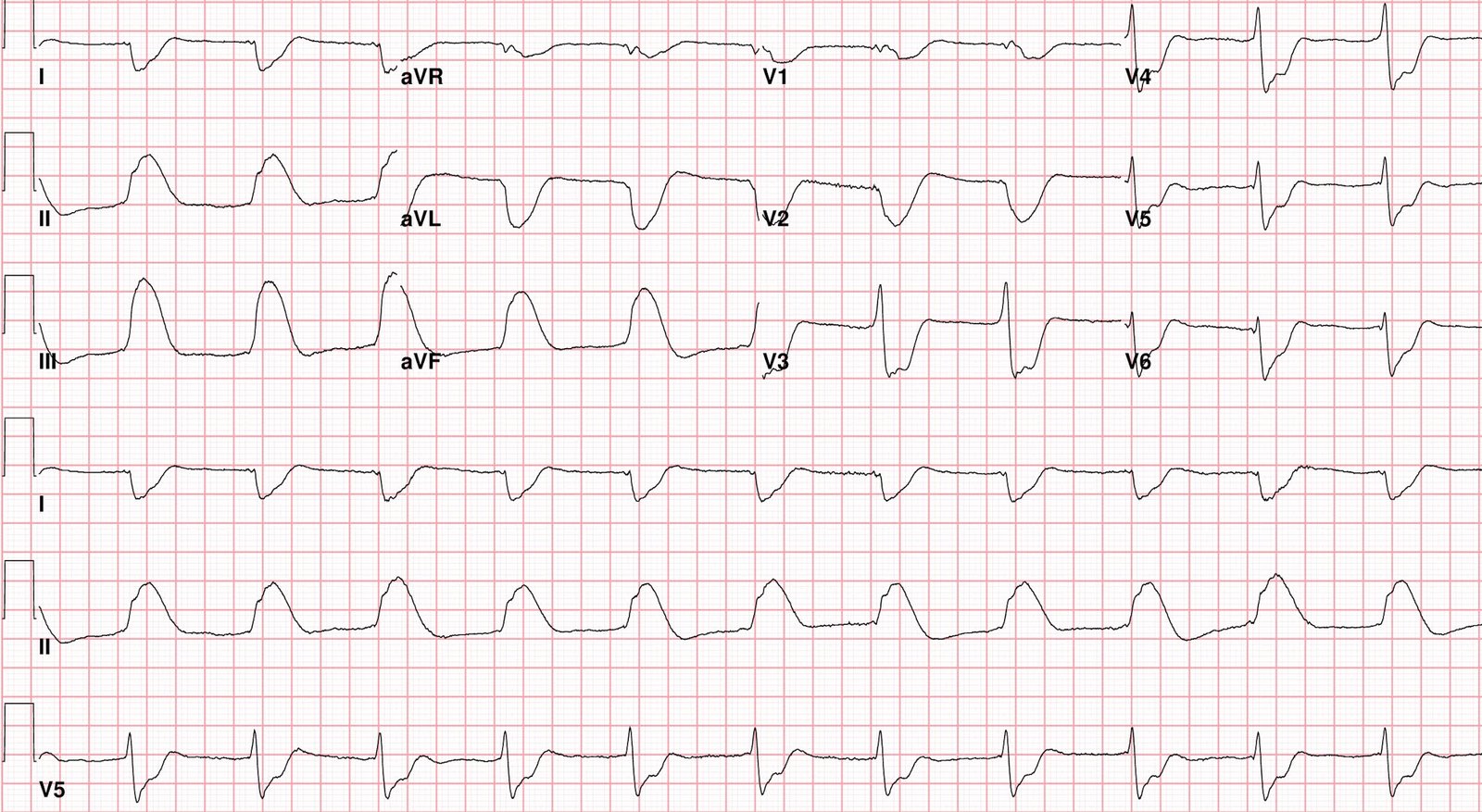
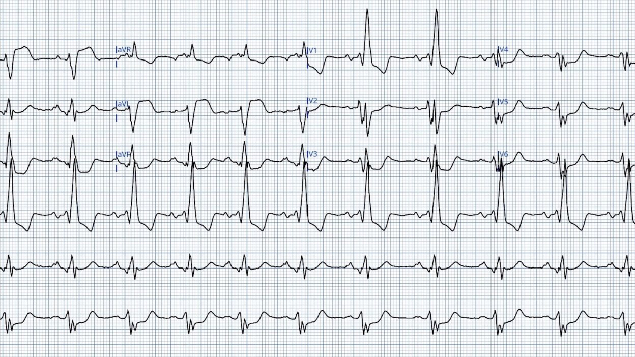
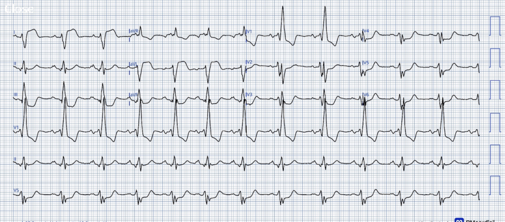
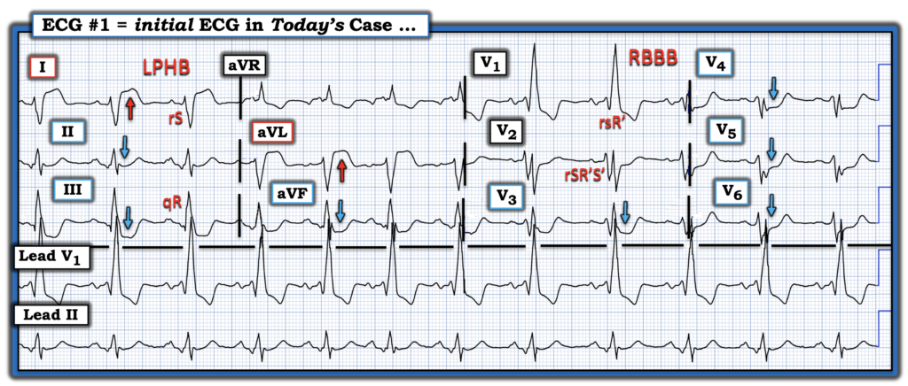
Smith: There is RBBB with Left posterior fascicular block (small inferior q-wave followed by large R-wave, and in aVL there is a small r-wave followed by a large S-wave. So: bifascicular block. Not as serious as RBBB + LAFB, but still it is bifascicular block. We don’t know if new or old. There is large ST Elevation in I and aVL. There is profound ST depression in V2-V6, maximal if V3. The ST depression in V2 is concordant to the S-wave of the RBBB. The ST depression in V3 is appropriately discordant to the R’-wave of RBBB, but it is out of proportion! So this looks like a circumflex occlusion with high lateral and posterior OMI. Other arteries can result in this territory of infarction.
The treating physician was not impressed.
His partner is one of my former residents, and he was impressed. He used the PMCardio Queen of Hearts and this is the result:
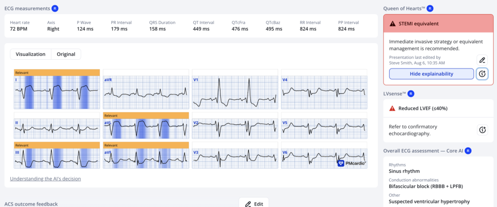
This convinced his partner to activate the cath lab.
Here is the result:
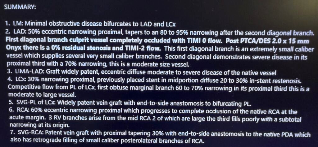
LAD: 50% eccentric narrowing proximal, tapers to an 80-95% narrowing after the 2nd diagonal. First diagonal is completely occluded with TIMI-0 flow.
Here is the entire cath report:
= = =
==================================
MY Comment, by KEN GRAUER, MD (8/7/2026):
Today’s case by Dr. Smith is a “feast” for the intellectually curious! I say this because I believe this case represents a unique but fascinating positive South African Flag Sign in a patient with a multi-fragmented bifascicular block.
= = =
Putting Today’s Case into Perspective:
There are 2 important aspects to today’s case:
- #1) = The Clinically Important Diagnostic Aspect: As per Dr. Smith’s former resident — Today’s initial ECG is an extremely worrisome tracing in this middle-aged woman with known coronary disease, who presents with new CP (Chest Pain). This tracing justifies the prompt cath lab activation that was ordered.
- #2) = The Fascinating Subtleties in this Initial ECG (See below).
= = =
The Initial ECG …
For clarity in Figure-1 — I’ve reproduced and labeled the initial tracing. As per Dr. Smith — the multiple abnormal findings in ECG #1 include the following:
- Sinus arrhythmia at a rate of ~70/minute with an obviously widened and multi-fragmented QRS complex. I interpreted the rSR’S’ in lead V2 (with similar-looking multi-fragmented complexes in the remaining chest leads) as “Q-wave-equivalents” from the scarring of prior infarction.
- There is bifascicular block in the form of RBBB/LPHB (as diagnosed by the rsR’ in V1 — and the rS/qR pattern in leads I and III, with a very deep S wave in lead I).
- Because the left posterior hemifascicle is much larger and thicker than the left anterior hemifascicle — it is far less common to see true LPHB, than it is to see LAHB.
- That said, when you do see true LPHB — it generally means there is extensive underlying structural disease — which is another reason why true LPHB is almost always accompanied by RBBB.
- As a result — I suspect the combination of RBBB/LPHB + multi-fragmented complexes in most of the 12 leads in Figure-1 are old findings from extensive prior infarction in a patient with severe underlying coronary disease (Finding a previous baseline ECG on this patient would be invaluable for confirming my suspicion).
- In contrast — the combination of RBBB/LAHB is much more commonly seen than RBBB/LPHB. When RBBB/LAHB is acute and occurs in association with anterior lead ST elevation — this form of bifascicular block tells us there is acute proximal LAD occlusion with worrisome acute conduction system damage.
- In the Chest Leads: There is dramatic (3-4 mm! ) ST elevation in leads I and aVL — with equally dramatic diffuse ST depression that is seen in virtually all other leads on this tracing.
- While difficult (impossible?) to determine “new” from “old” from “new-superimposed-on-old” from this single ECG — this amount of ST elevation and ST depression in this patient who now presents with new CP — has to be viewed as an acute OMI in progress until proven otherwise (At this point in the process — determining the acute “culprit” lesion is an academic exercise that pales in the light of knowing that prompt cath lab activation is needed!).
= = =
Figure-1: The initial ECG in today’s case. (To improve visualization — I’ve digitized the original ECG using PMcardio).
= = =
South African Flag + Bifascicular Block
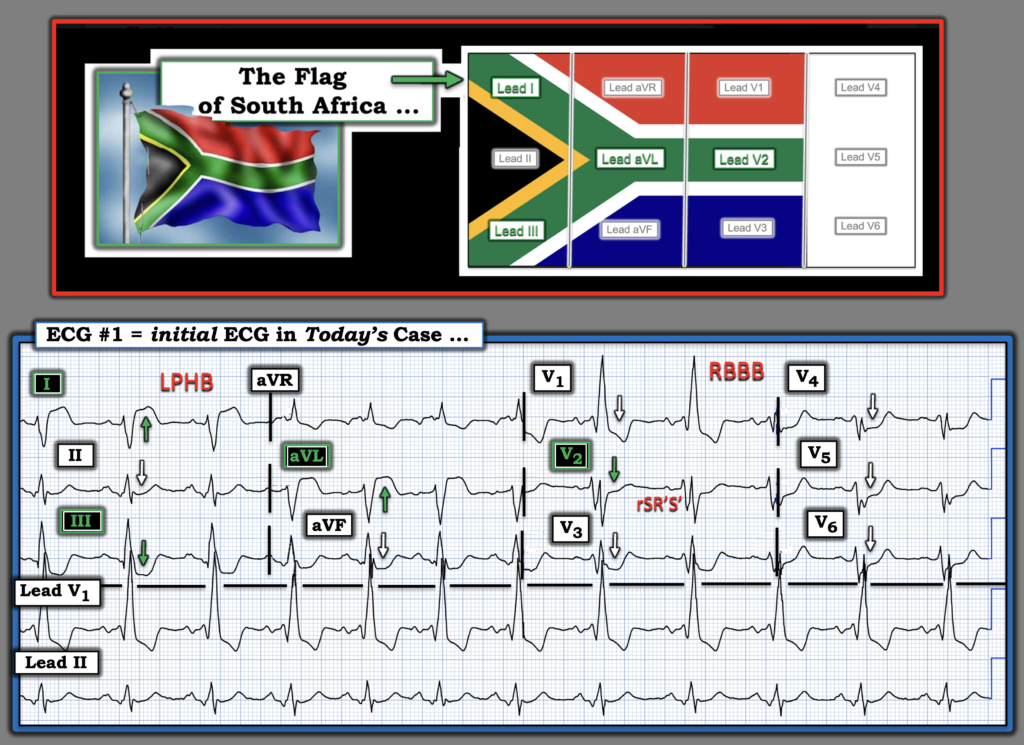
With full realization that highest priority in today’s case is expedition of cardiac cath with PCI — I was intellectually curious as to Why there was relatively little ST depression in lead V2 compared to the 5 other chest leads?
- My Suspicion: This initial ECG represents a South African Flag Sign in this patient with bifascicular block and severe underlying coronary disease.
We have shown many cases in Dr. Smith’s ECG Blog of the South African Flag pattern — as an indication of acute occlusion of the 1st or 2nd Diagonal Branch of the LAD.
- As highlighted in the top portion of Figure-2 — the S. Afr. Flag pattern is recognized by the findings of:
- ST elevation in leads I and aVL;
- Reciprocal ST depression in lead III;
- ST elevation in lead V2 — but not in any of the other chest leads (which typically show ST depression).
While none of the chest leads in Figure-2 manifest ST elevation — I found lead V2 remarkable for its paucity of ST depression compared to the other 5 chest leads.
- I suspect the reason for this relative lack of ST depression in lead V2 — is simply that the ST elevation that would have been seen in lead V2 from this patient’s cath-confirmed acute 1st Diagonal occlusion — has been attenuated by the marked ST depression that results from the combined effect of this patient’s RBBB and severe diffuse subendocardial ischemia.
= = =
Figure-2: I see a positive South African Flag Sign.
= = =
= = =