Written by Magnus Nossen .
This ECG is from a man in his early 50s with a history of bilateral pulmonary embolism who presented with acute shortness of breath and chest pain. How would you interpret the ECG?
ECG #1
What do you think?
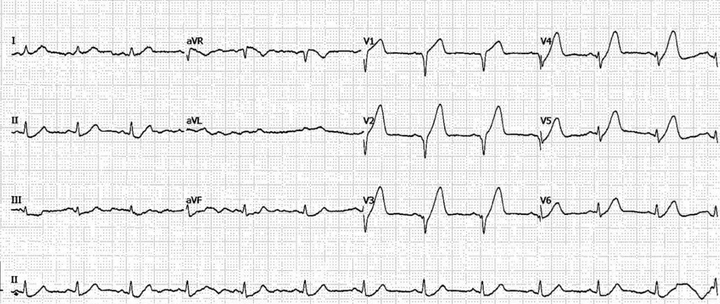
This ECG shows sinus rhythm 74 beats per minute. QRS duration is normal. There is loss of R-wave in leads V1-V3, with no more than a minimal R wave in lead V4. The J point is depressed in leads V3-V6, with a steep ascending ST segment that leads into hyperacute T waves. This pattern is absolutely diagnostic of LAD OMI (referred to in the literature as “deWinter T waves”).
Here is the PMCardio Queen of Hearts AI ECG Model interpretation:
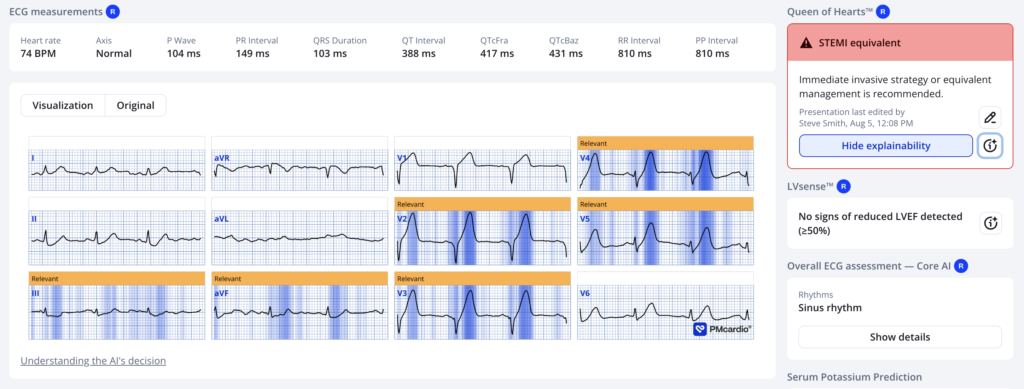
The numerical output was 1.00 (as high a confidence as is possible)
To readers of this blog — the pattern in ECG #1 is instantly recognizable as LAD OMI.
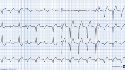
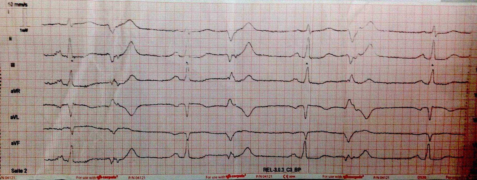
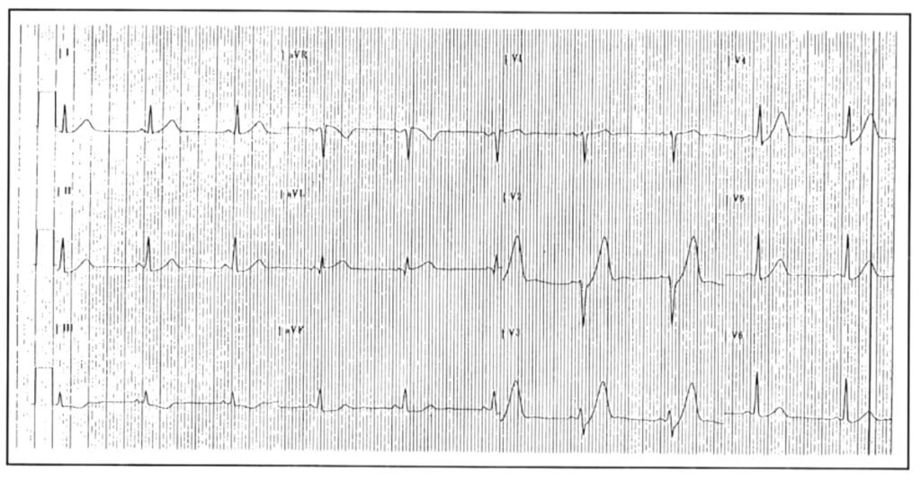
- Of historical note — this finding of tall precordial T waves with depressed J point was initially described by CS Soo in a 1995 case report (and not by deWinter).
- Below is the ECG from this original 1995 case report (Soo CS — Singapore Med J: 36:216, 236-237, 1995).
Back to the Case:
The computer algorithm interpreted ECG #1 as showing, “Normal sinus rhythm, low voltage QRS; cannot rule out anteroseptal infarct, age undetermined”.
- From hospital arrival (and the recording of ECG #1) until cath lab activation — 60 minutes passed. This is too long for a patient with an instantly diagnostic ECG. The delay likely reflects failure to recognize ECG #1 as a STEMI equivalent.
- The Queen of Hearts AI model identified ECG #1 as OMI, as shown above.
The coronary angiogram showed, “LAD 90% stenosis; LCx 50% stenosis; OM1 99% stenosis”.
- TIMI 3 was noted in all coronaries, and it was decided the patient was best managed with CABG surgery.
- Initial conventional Troponin I returned at 27ng/L (ref < 40ng/L).
- No PCI was done at this time.
Following cardiac cath, while arrangements for transfer to a facility with cardiothoracic surgery were being made — ECG #2 was recorded.
- This repeat ECG shows significant change when compared to the admission ECG.
- What is going on? What is your management?
= = =
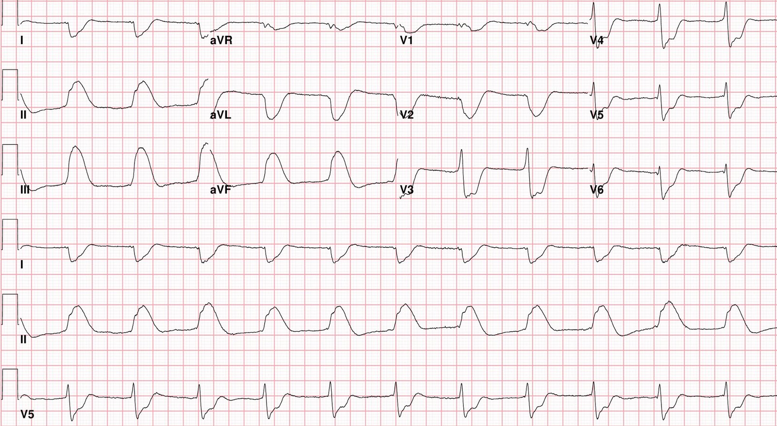
ECG #2
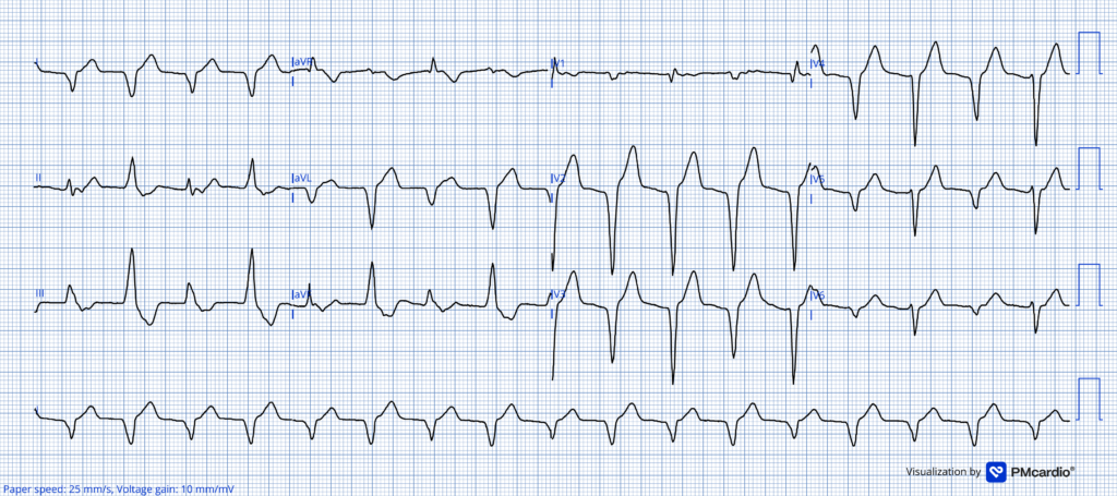
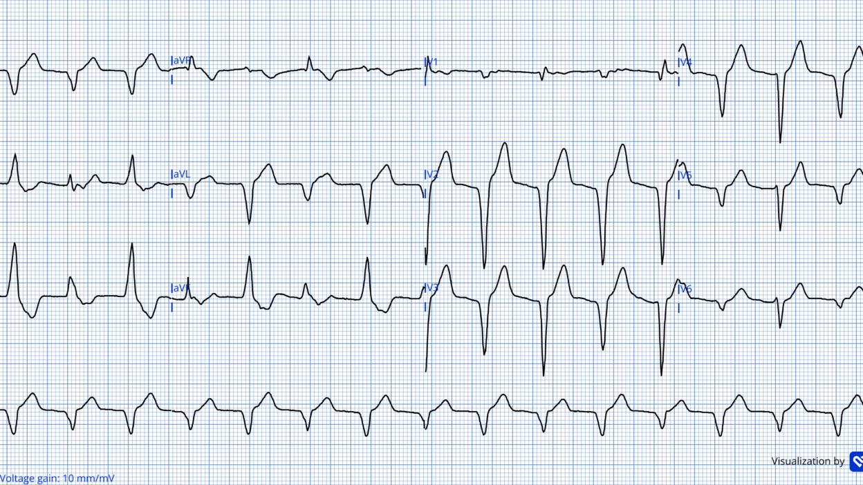
ECG #2 shows a regular wide complex rhythm with a rate of 103 beats per minute. QRS complexes are entirely negative in leads I and aVL, and predominantly negative in the chest leads. There is beat-to beat alteration of QRS morphology. This is accompanied by slight axis change, but not to the extent seen in bidirectional VT in which the QRS axis usually alternates 180 degrees. (For a review of bidirectional VT have a look at this case, this case and this case).
= = =
ECG #2 is consistent with AIVR (Accelerated IdioVentricular Rhythm) with 2 foci of automaticity. AIVR is almost always unifocal — but in ECG #2 there are clearly 2 alternating foci of automaticity accounting for the 2 distinct QRS morphologies.
- AIVR is recognized by its regular wide complex without a preceding P wave and comparatively slow rate compared to VT
- AIVR is usually well tolerated and rarely requires intervention. In most cases, the key is recognizing that AIVR is present and knowing that it almost never needs active treatment.
- AIVR is a common reperfusion arrhythmia, typically occurring at a rate of 60–110 beats per minute. This rhythm is usually transient, often lasting only minutes and, more rarely, hours to days. It is caused by increased automaticity and is usually unifocal, unlike the rhythm seen in this case.
- Overall, AIVR is a favorable prognostic sign that suggests there has been coronary reperfusion.
Clinically — today’s patient was hemodynamically stable at the time ECG #2 was seen. The patient’s chest pain had resolved.
- No specific treatment was given for the bifocal AIVR in today’s case, which spontaneously resolved after a limited period of time.
- The Queen of Hearts AI model did not recognize this example of AIVR as a reperfusion ECG.
= = =
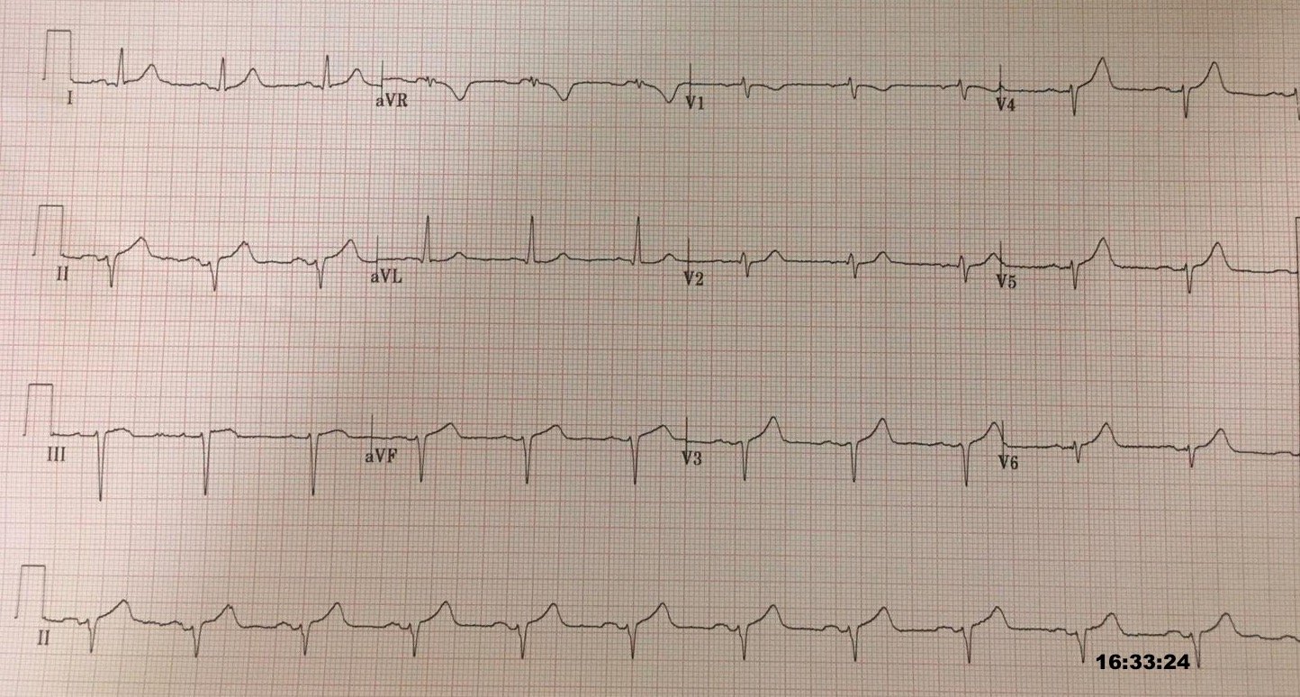
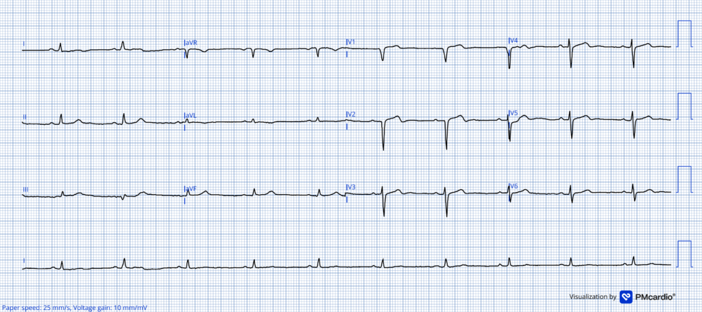
ECG #3 was recorded about 12 hours after admission and just prior to transfer for CABG surgery. AIVR did not recur — and normal sinus rhythm was established. Note that signs of reperfusion are now evident in ECG #3 by the presence of subtle anterior reperfusion T waves.
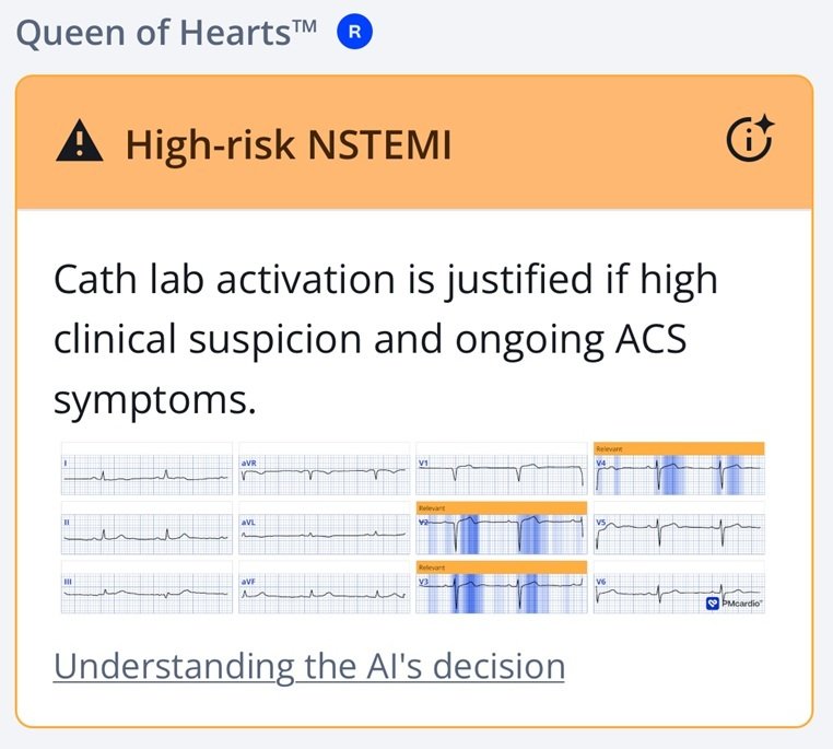
- As shown below — reperfusion patterns are classified as high risk NSTEMI by the Queen of Hearts.
- Troponin I rose to 3960ng/L but was not measured to peak.
- Long term follow up for this patient is unavailable.
ECG #3
= = =
Learning Points:
- With a diagnostic ECG (as was seen in ECG #1) — the time from arrival to the cath lab should be significantly shorter than 60 minutes.
- AIVR that occurs in the setting of acute OMI is a reperfusion arrhythmia. In most instances — no treatment is needed.
- AIVR is almost always unifocal and monomorphic. However, as illustrated by today’s case — AIVR can manifest alternating QRS morphologies.
= = =
==================================
MY Comment, by KEN GRAUER, MD (7/31/2026):
I found today’s case of interest for several reasons:
- It highlights a number of important features of today’s deWinter T wave pattern — with the frustrating reality that even when deWinter T waves are clearly diagnostic, this pattern still goes unrecognized (as it initially was in today’s case).
- It raises questions as to when is it safe to delay PCI reperfusion in a patient with multivessel disease scheduled for CABG the next day?
- It teaches us about a rare but very important arrhythmia that we need to be aware of.
= = =
Today’s initial ECG …
As per Dr. Nossen, today’s initial ECG (that I have reproduced in Figure-1) — should have been recognized as definitive enough to prompt immediate activation of the cath lab.
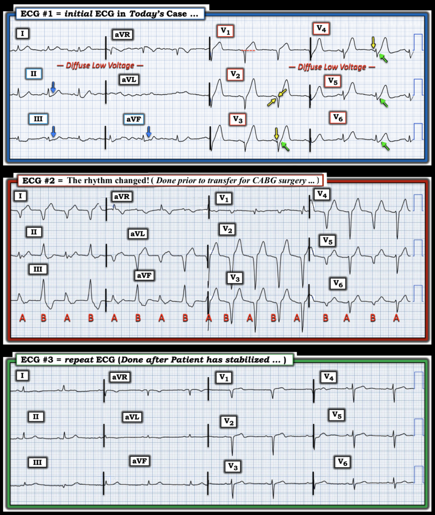
- Classic deWinter T waves are present in leads V2-thru-V6 of today’s initial tracing. By this I mean that beginning with J-point ST depression (GREEN arrows in ECG #1 ) — the ascending T wave limb manifests a steep incline that culminates in giant T wave (that attains 10 mm in height in leads V3,V4) — with a “fat” T wave peak and a wider-than-expected T wave base.
- Note that the shape of the hyperacute T wave in lead V1 is different that the shape of the deWinter waves in leads V2-thru-V5. Instead of the concave-up steeply ascending T waves that are seen in the rest of the chest leads — there is straightening of the ST segment takeoff of this overly “bulky” (hyperacute) T wave in lead V1 that also manifests J-point ST elevation. This strongly suggests a very proximal LAD lesion.
- Reciprocal ST depression in each of the inferior leads (BLUE arrows in leads II,III,aVF) — provides additional support of a more dangerous proximal LAD lesion.
- Note the diffuse Low Voltage (present in all 12 leads). As we’ve often emphasized in Dr. Smith’s ECG Blog — among the many potential causes of low voltage is a large acute infarction with cardiac “stunning” (which should alert providers to the possibility of impending cardiogenic shock — See the August 3, 2025 post for more on the causes and implications of low voltage).
- Finally — Not only are there QS waves in leads V1,V2,V3 — but the initial slurring on the downslope of the S wave in leads V2,V3 (YELLOW arrows in ECG #1) — in association with the small initial q wave in lead V4 and the clearly diminished R wave amplitude in leads V4,V5,V6 — establishes that there has been extensive anterior infarction at some point in time.
My Clinical Impression of the initial ECG:
- An extensive acute infarction is clearly ongoing in today’s case! Simply by looking at ECG #1 and being aware of the history we were provided with (ie, of a man in his 50s who presented with chest pain and acute shortness of breath) — I fully expected cath results to indicate acute proximal LAD occlusion.
- Whether there had been a previous anterior infarction, now with superimposed new LAD occlusion — or whether all chest lead findings in ECG #1 reflected a single extensive ongoing acute event was uncertain from this limited history and lack of a prior tracing.
- Cath results provided by Dr. Nossen revealed severe multi-vessel disease without specifying the acute “culprit” vessel — and, with the surprising finding that the most severe lesion was not the LAD, but the 99% narrowing of OM-1.
- Again, in the setting of the above noted findings — the diffuse low voltage raises concern of cardiac “stunning” with severely impaired LV function.
= = =
Figure-1: Serial tracings of the 3 ECGs in today’s case.
= = =
And then the Rhythm Changed …
As noted above in Dr. Nossen’s discussion, despite severe multi-vessel disease demonstrated on cath — the initial Troponin was normal, and the patient appeared hemodynamically stable. As a result — the decision was made not to perform PCI, but instead to transfer the patient to a facility with CABG capability. And then the rhythm changed … (as shown in ECG #2).
- As described by Dr. Nossen — ECG #2 manifests 2 distinct QRS morphologies that switch back-and-forth with every beat ( = alternating complexes labeled “A”and “B” ). Both morphologies feature a wide QRS. P waves are absent. The R-R interval between complexes appears to be relatively constant at a heart rate close to 100/minute.
- Impression: This is clearly a ventricular rhythm with 2 different QRS morphologies that alternate every-other-beat.
My differential diagnosis for the rhythm in ECG #2 consisted of 3 possible entities:
- i) AIVR with 2 ventricular foci (I’ll call this “bifocal AIVR” ).
- ii) Bidirectional VT.
- iii) AIVR from a single ventircular focus, but with alternating exit sites or beat-to-beat variation of the ectopic reentry circuit.
We’ve presented examples of BiDirectional VT (See My Comment at the bottom of the page in the February 18, 2026 post, among others). In many ways — bifocal AIVR and bidirectional VT look similar in that they are each exclusive ventricular rhythms that manifest 2 alternating QRS morphologies. Theoretically — the differences between these 2 entities include the following:
- Bidirectional VT tends to be a faster VT form with stricter alternation of the QRS axis that typically flips ~180 degrees from one beat-to-the-next (often simulating alternation of lbbb and rbbb conduction patterns). The alternating pattern of QRS morphologies with bidirectional VT often manifests alternating longer-then-shorter R-R intervals.
- In contrast — bifocal AIVR tends to be a slower ventricular rhythm form that manifests a more regular R-R interval with less extreme axis shift between the 2 ventricular morphologies. There is less consistency in beat-to-beat variation — such that one QRS morphology may take over for a period of time before switching back to the other QRS morphology. And there may be more than 2 different QRS morphologies that are seen.
- Complicating distinction between bidirectional VT vs bifocal AIVR — is the reality alternating QRS morphologies in a ventricular rhythm may be the result of duplicate (or multiple) exit sites, or duplicate VT “circuits”, or shifting conduction properties within a given circuit that alters the activation sequence (See My Comment in the June 1, 2020 post).
Bottom Line ( = My Thought) — It may not always be easy to distinguish between bidirectional VT vs bifocal AIVR, as there may be overlap between the 2 entities.
- Clinically — The significance of which VT form is present relates to the expected outcome. Both bidirectional VT and bifocal AIVR may occur in association with acute ischemia/OMI.
- Bifocal AIVR tends to behave clinically like the much more common uniform AIVR — which almost always serves as a “good” sign that there has been reperfusion of the “culprit” vessel. In this setting — AIVR (uniform or bifocal) is most often a transient rhythm that requires no treatment, because it will almost always “go away” within minutes or a few hours.
- In contrast — bidirectional VT that arises as a result of acute ischemia is a much more dangerous rhythm at risk of deteriorating to VFib.
= = =
Application of ECG #2 to Today’s Case:
Dr Nossen in his discussion conveyed the “good news” in today’s case.
- Features suggesting that the rhythm in ECG #2 was bifocal AIVR (and not bidirectional VT) — were that the overall ventricular rate was slower, the axis shift between the 2 ventricular morphologies was less extreme — and the patient was hemodynamically stable.
- As a result — the decision was made not to treat the rhythm in any way, and sinus rhythm return within a short period of time.
= = =
The Final ECG in Today’s Case …
As per Dr. Nossen — ECG #3 was recorded ~12 hours after ECG #1, just prior to transfer of the patient to the CABG-capable facility.
- Going back to Figure-1 — We see deflation of virtually all acute ST-T wave changes that had been present in ECG #1.
- There are ECG signs that even without PCI — some element of spontaneous reperfusion has occurred (ie, beginning T wave inversion is now seen in leads aVL and V2, if not also in other chest leads).
- Additional promising ECG findings in ECG #3 include return of an initial R wave in leads V3,V4 — and slight increase in QRS amplitude in lead V2-thru-V6.
= = =
= = =