Written by Magnus Nossen.
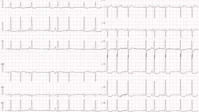
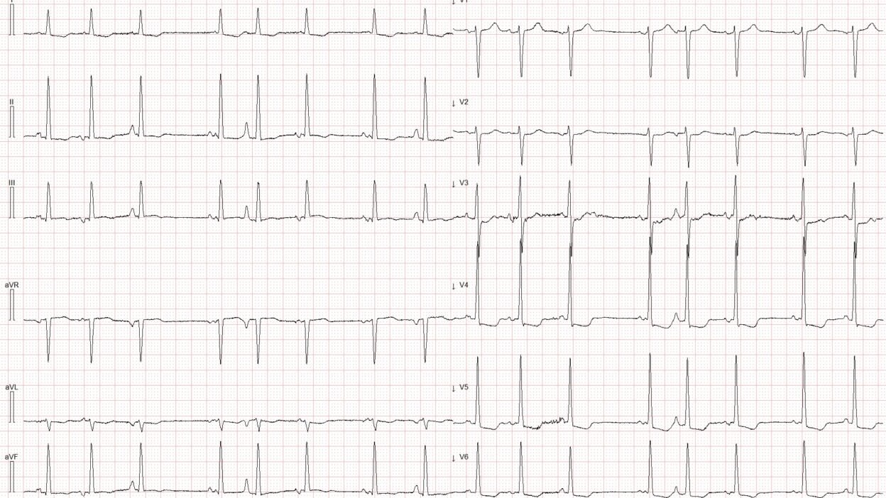
An ECG was obtained from an elderly male nursing home resident who was referred to the emergency department because of shortness of breath and confusion. His past medical history includes Alzheimer’s disease, hypertension, and COPD. He was clinically stable on arrival. How would you interpret the ECG shown?
ECG #1
Smith: What do you think?
Is there subendocardial ischemia?
Is there OMI and subenendocardial ischemia (SEI)?
Is this only LVH?
Is it both LVH and subendocardial ischemia?
Are there all 3: LVH and SEI and OMI?
Magnus continues:
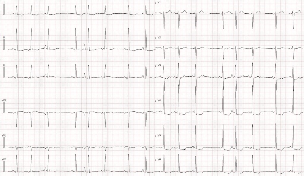
This ECG demonstrates an irregularly irregular rhythm.
99% of the time, an irregularly irregular rhythm is atrial fibrillation. So, is it atrial fibrillation?
More importantly: There is voltage evidence of LVH and ST‑segment depression in leads I, aVL and V3–V6. The ST segment in lead III varies some with P‑wave morphology — but is overall isoelectric, with perhaps a subtle hint of minimal ST elevation in some beats.
Together, these ST-T changes in the setting of new onset dyspnea is worrisome for ongoing ischemia. Sometimes it can be difficult to discern subendocardial ischemia (SEI) from OMI — and sometimes the patterns overlap.
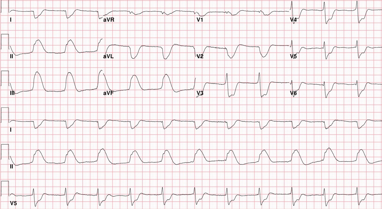
In particular I find it difficult to clearly distinguish Aslanger’s pattern from SEI. The below image shows how the ST vector of Aslangers pattern lies between the ST vector of SEI and inferior OMI. This explains why these patterns may resemble each other.
Figure #1:
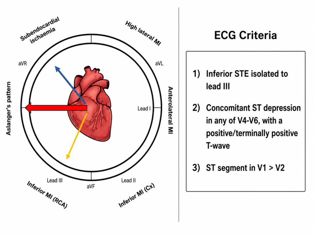
Figure 1 show the ST segment vector (left) and the ECG criteria (right) for Aslanger’s pattern. Figure adapted from Dr Smith and Life in the Fast Lane.
= = =
ECG# 1 lacks terminally positive T waves in V4-V6 and there is no clear ST elevation in lead III meeting official criteria. As such, this ECG resembles the subendocardial ischemia pattern more than Aslanger’s pattern. I think it is likely there is SEI superimposed on left ventricular hypertrophy causing the ST-T changes.
The QoH AI model correctly flagged these ST-T changes as concerning for ischemia, but does not say OMI. The AI model recommended serial ECGs to monitor for dynamic changes and evolution toward overt OMI. Serial ECGs were not performed; neither was bedside ECHO.
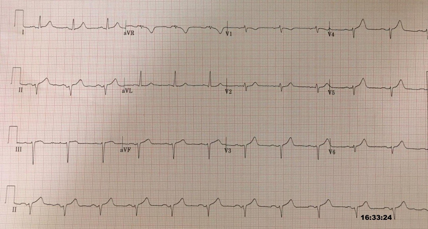
Figure #2:
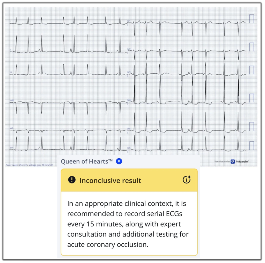
The Queen of Hearts AI model classified ECG #1 as “Inconclusive.”
Smith: our experimental model (which names OMI equivalents and mimics, but is not yet available) gives a result of:
1) Subendocardial ischemia 66% and 2) LVH 68%, but not OMI.
This indicates that none of the thresholds for active OMI or reperfused OMI, were met — although one may have been near its respective diagnostic cutoff. Given the clinical context in today’s case — this classification represents a high-risk scenario, such that the model recommends close clinical monitoring and further diagnostic evaluation.
Due to severe dementia the patient was unable to provide a reliable history, but he denied chest pain. The absence of chest pain does not of course rule out ischemia. The patient was admitted with a working diagnosis of NSTEMI after the initial hs‑cTnI returned at 75 ng/L. The subsequent hs‑cTnI rose dramatically to 56,420 ng/L, a more compatible with OMI than SEI. No further troponin measurements were obtained. Clinically, the patient developed progressively worsening dyspnea, and the decision was ultimately made to transition to comfort‑focused care.
Smith: SEI generally has hs-cTnI well below 5000 ng/L, so I suspect that this really was an acute coronary occlusion (OMI). OMI can be so subtle on the ECG as to show nothing. This patient has definite refractory ischemia (whether SEI or OMI does not matter!). It is classified as refractory because the ischemic ST changes are not resolving with medical therapy. Refractory ischemia needs emergent cath lab activation regardless of OMI or SEI. That is why the European guidelines recommend emergent angiography for “NSTEMI” with a very high suspicion “regardless of ECG or biomarker findings.”
Smith: The acute dyspnea certainly could be due to acute ischemia, though the data given is not sufficient to give us an explanation for the symptoms.
In this particular case, recognizing the ECG changes would probably not have changed management, as this elderly man was not a candidate for coronary angiography. That said — clinical implications of the above findings might have been very different in a patient with better baseline functional status.
The ST-T changes in today’s case are interesting in their own right. But did you notice the type of rhythm present on todays ECG? If not — Have another look at ECG #1 above.
= = =
The rhythm in ECG #1 is irregularly irregular with P waves of varying morphology. I can identify at least 5 distinct P‑wave morphologies in this ECG. The most common explanation for changing P‑wave morphology is atrial ectopy. When there are 3 or more different P‑wave morphologies in the setting of an irregularly irregular rhythm and the heart rate exceeds 100 bpm — this is termed multifocal atrial tachycardia or “MAT”. In today’s tracing, however, the ventricular rate is below 100 bpm.
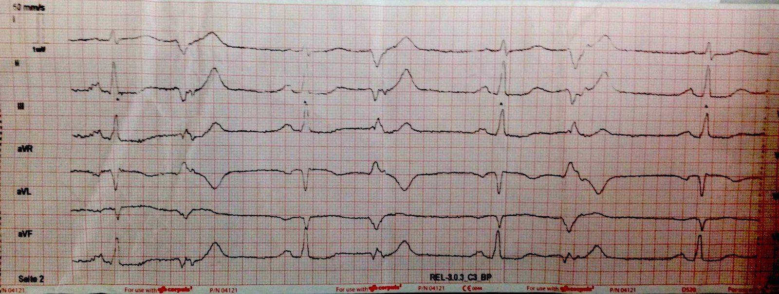
Figure #3:
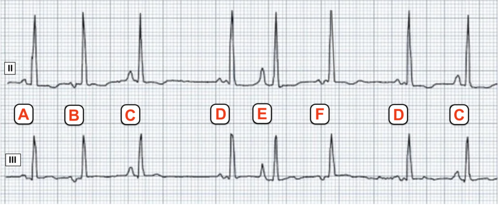
In this Figure #3 — I have reproduced lead II (top) and lead III (bottom) from ECG #1. Different P wave morphologies are labeled with letters A-F. (Note that P wave morphologies C and D appear twice. It’s possible that morphologies B and F may be from the same atrial focus).
The rhythm in today’s case would meet diagnostic criteria for multifocal atrial tachycardia (MAT) if the heart rate was slightly faster. MAT is defined as an irregularly irregular supraventricular rhythm with a rate greater than 100 beats per minute with at least 3 distinct P-wave morphologies, and one P wave for each QRS complex. It is commonly associated with significant pulmonary disease and/or hypoxia. The threshold of 100 beats per minute is arbitrary and is not related to the underlying precipitating factors. Our patient may not satisfy the formal heart rate criterion for MAT. It is reasonable to describe this as Multifocal Ectopic Atrial Rhythm, which can be viewed as part of the same spectrum as MAT, but with a heart rate below 100 beats per minute.
In younger, otherwise healthy individuals, one may occationally observe a wandering atrial pacemaker (WAP). This is typically regarded as a benign rhythm associated with low resting sinus rates and enhanced vagal tone. In true WAP, P wave morphology changes gradually. This is unlike the rhythm in ECG #1 where P wave morphology is changing dramatically from beat to beat.
Final Thought: I suspect the rhythm in today’s case is not directly related to the ongoing ischemia — but is more likely incidental and related to the underlying COPD this patient was known to have.
(See Ken Grauer discussion of the rhythm in his Comment below.)
= = =
Learning Points:
- SEI and OMI can be difficult to distinguish on the ECG alone, and there is often some overlap between these two entities.
- High risk ECG patterns need close monitoring and clinical investigation.
- MAT is typically seen in patients with pulmonary disease and/or hypoxia, and the changes in P-wave morphology are abrupt.
- WAP is usually a benign rhythm seen in otherwise healthy individuals. As opposed to MAT — the changes in P-wave morphology with WAP are gradual.
= = =
==================================
MY Comment, by KEN GRAUER, MD (7/28/2026):
I focus My Comment on the rhythm in today’s case. As per Dr. Nossen — this patient presents in an irregular rhythm with an overall rate of less than 100/minute, and with P waves of varying morphology.
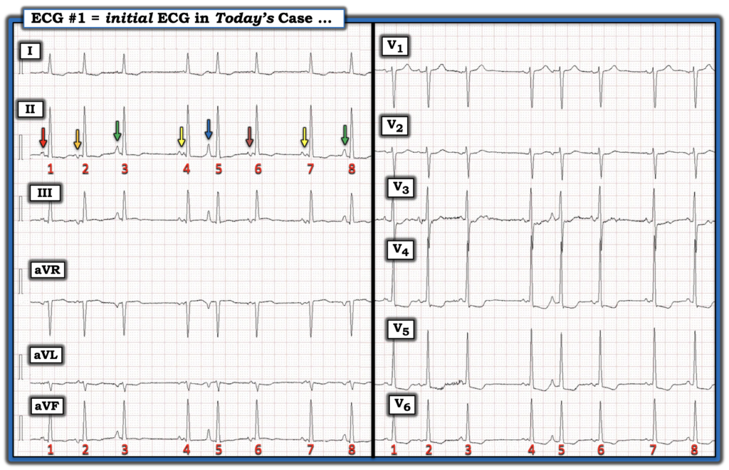
- For clarity in Figure-4 — I’ve numbered the beats and added colored arrows to highlight the varying P wave shapes in today’s initial ECG.
= = =
The Rhythm in Figure-4 …
We’ve addressed the entity of MAT (Multifocal Atrial Tachycardia) on a number of occasions in Dr. Smith’s ECG Blog (See My Comment in the January 5, 2020 — and September 30, 2019 posts, among others).
- To emphasize — The same 8 beats are shown in the limb and chest leads. It is difficult to express certainty about a rhythm with a sample limited to 8 beats.
- The P waves preceding beats #3 and 8; and preceding beats #4 and 7 appear to arise from the same atrial focus (duplicate GREEN and YELLOW arrows, respectively).
- The other 4 P waves all clearly differ in morphology.
- I have no idea which of the 6 different P wave shapes in Figure-4 represents the “sinus” P wave in this irregular rhythm without pattern.
= = =
The usual differential diagnosis to consider when one sees an irregular supraventricular rhythm with variable P wave morphology includes: i) Sinus rhythm with multiple PACs; — ii) WAP (Wandering Atrial Pacemaker); — and, iii) MAT.
- The rhythm in Figure-4 is too irregular and lacking in a recognizable sinus P wave shape to be “sinus rhythm” with multiple PACs.
- This rhythm is also not WAP — because change in the atrial pacing site is gradual with that disorder (as per My Comment at the bottom of the page in the May 19, 2022 post). In contrast — we never see the same P wave morphology in consecutive beats in Figure-4 (See the ADDENDUM below for more on wandering pacemaker).
- Instead, given the change in P wave morphology and PR interval from one-beat-to-the-next — this would seem to suggest MAT as the rhythm in today’s case — except that the overall ventricular rate is not tachycardic!
- Some cardiac rhythms do not read the textbook! Having observed this phenomenon over decades — I’ve noticed that rather than black-or-white classification for rhythms such as wandering pacemaker; sinus rhythm with many PACs; and MAT — that there is a spectrum for these rhythm disorders. (CLICK HERE — for my detailed discussion of this subject).
- Clinically — the rhythm we see in today’s tracing behaves similar to MAT, even though there is no tachycardia. A majority of patients with MAT have longstanding pulmonary disease. Rather than antiarrhythmic medication — optimizing pulmonary function is the best treatment approach. Features in today’s case consistent with my conclusion regarding today’s rhythm include the following:
- Today’s patient has a history of COPD — and his presenting symptom is shortness of breath.
- Among the P wave shapes in Figure-4 — are the pointed P waves of RAA (preceding beats #3,5,8) — with the BLUE arrow P wave preceding beat #5 being extremely tall and consistent with the telltale sign of significant underlying pulmonary disease.
= = =
Final Thought: Practically speaking — it is both academic and clinically unimportant (as well as often impossible) to attempt to distinguish between sinus rhythm with multiple different-looking PACs vs MAT. This is because:
- i) The same predisposing clinical scenario is common to both of these arrhythmias; — and,
- ii) Optimal management (ie, To identify and treat the underlying cause of the arrhythmia) is the same for both conditions.
= = =
Figure-4: The initial ECG in today’s case.
= = =
ADDENDUM (re MAT vs Wandering Pacemaker):
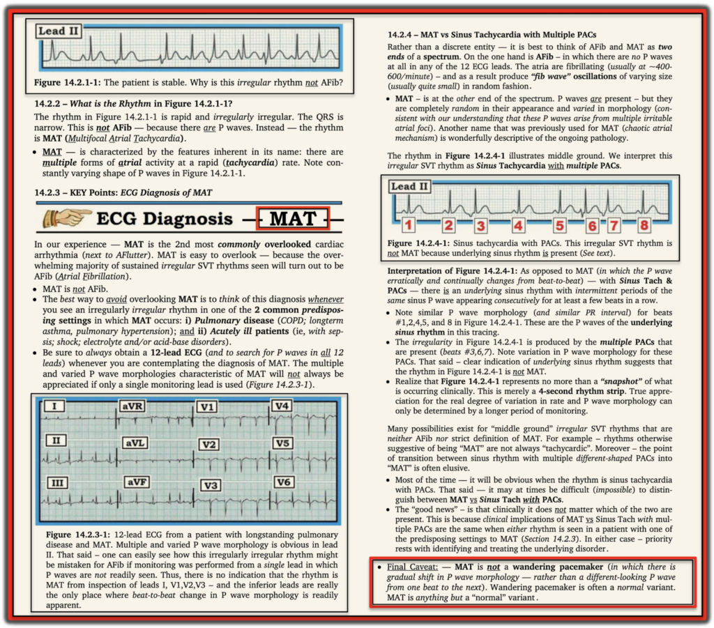
- 3:25 minute Video Pearl on MAT.
- 1-Page Summary of MAT (from Grauer K: 2013 ACLS/Arrhythmia ePub).
= = =
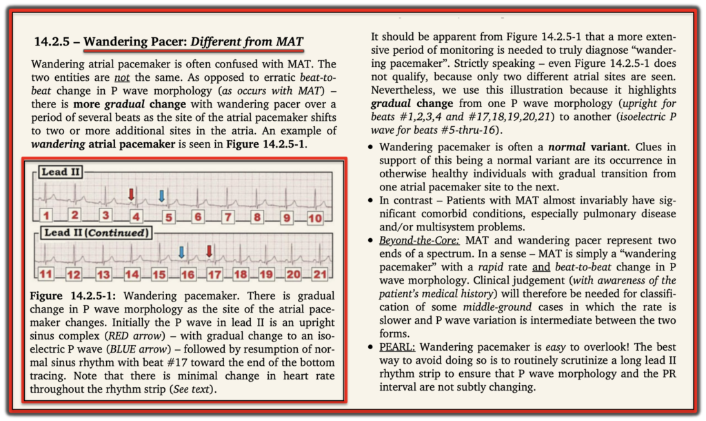
- 3:30 minute Audio Pearl on Wandering Pacemaker.
- 1-Page Summary of Wandering Pacemaker (from Grauer K: 2013 ACLS/Arrhythmia ePub).
= = =
= = =